A study by the Universities of Sussex and Portsmouth reveals that horses can read and then remember people's emotional expressions, enabling them to use this information to identify people who could pose a potential threat.
Published today, Thursday 26 April 2018, in the journal, Current Biology, the paper 'Animals remember previous facial expressions that specific humans have exhibited' is authored by a team of psychologists, co-led by Professor Karen McComb from the University of Sussex and Dr Leanne Proops, from the University of Portsmouth -- both specialists in animal behaviour.
The research team conducted controlled experiments in which domestic horses were presented with a photograph of an angry or happy human face and several hours later saw the actual person who had exhibited the expression, now in an emotionally neutral state. This short-term exposure to the photograph of a person's facial expression was enough to generate clear differences in subsequent responses upon meeting that individual in the flesh later the same day.
The study found that despite the humans being in a neutral state during the live meeting, the horses' gaze direction revealed that they perceived the person more negatively if they had previously seen them looking angry in the photograph rather than happy. Previous research, including at University of Sussex, has shown that animals tend to view negative events with their left eye due to the right brain hemisphere's specialisation for processing threatening stimuli (information from the left eye is processed in the right hemisphere).
Importantly, in the current experiment the humans did not know which photographs the horses had previously seen, to avoid any risk of behaving differently themselves. Also the differences in reaction only applied to the person the horses had actually seen in the photograph and were not given to a different person.
Professor Karen McComb from University of Sussex comments on the findings: "What we've found is that horses can not only read human facial expressions but they can also remember a person's previous emotional state when they meet them later that day -- and, crucially, that they adapt their behaviour accordingly. Essentially horses have a memory for emotion."
Co-lead author Dr Leanne Proops, of the University of Portsmouth, said: "We know that horses are socially intelligent animals, but this is the first time any mammal has been shown to have this particular ability. What's very striking is that this happened after just briefly viewing a photograph of the person with a particular emotional expression -- they did not have a strongly positive or negative experience with the person."
Read more at Science Daily
Apr 29, 2018
Apr 28, 2018
Why a robot can't yet outjump a flea
![]() |
| The award for the fastest punch goes to mantis shrimp, which use their hammer-like appendages to smash open snail shells for food. |
New research could help explain why nature still beats robots, and describes how machines might take the lead.
Take the smashing mantis shrimp, a small crustacean not much bigger than a thumb. Its hammer-like mouthparts can repeatedly deliver 69-mile-per-hour wallops more than 100 times faster than the blink of an eye to break open hard snail shells.
Or the unassuming trap-jaw ant: In a zero-to-60 matchup, even the fastest dragster would have little chance against its snapping mandibles, which reach speeds of more than 140 miles per hour in less than a millisecond to nab their prey.
One of the fastest accelerations known on Earth is the hydra's sting. These soft-bodied aquatic creatures defend themselves with help from capsules along their tentacles that act like pressurized balloons. When triggered, they fire a barrage of microscopic poison spears that briefly accelerate 100 times faster than a bullet.
In a study to appear April 27 in the journal Science, researchers describe a new mathematical model that could help explain how these and other tiny organisms generate their powerful strikes, chomps, jumps and punches. The model could also suggest ways to design small, nature-inspired robots that come closer to their biological counterparts in terms of power or speed.
The secret to these organisms' explosive movements isn't powerful muscles, but rather spring-loaded parts they can cock and release like an archer's bow, said Sheila Patek, associate professor of biology at Duke University.
Tough yet flexible tendons, cuticles and other elastic structures stretch and release like slingshots, powering their jumps and snaps.
A short-legged insect called the froghopper, for example, has a bow-like structure called the pleural arch that acts like a spring. Latch-like protrusions on their legs control its release, allowing them to leap more than 100 times their body length despite their short legs. A person with that much power could jump nearly two football fields.
However, it's not clear how these mechanisms work together to enhance power, said Mark Ilton, a postdoctoral fellow at the University of Massachusetts Amherst.
While traditional mathematical models of performance take into account the inherent physical tradeoffs of muscle -- which can contract forcefully, or quickly, but not both -- they fail to factor in the tradeoffs inherent to springs and latch-like mechanisms as well. In other words, nothing can be faster, stronger, and more powerful at the same time.
"Until now these other components have been mostly black-boxed," Patek said.
The researchers developed a mathematical model of fast motion at small scales that incorporates constraints on springs and latches.
"Part of our goal was to try to develop a model that is equally generalizable to biological or engineered systems," said Manny Azizi, an assistant professor of ecology and evolutionary biology at the University of California, Irvine who studies jumping frogs.
First, they compiled data on the size and top speeds and accelerations for 104 species of elite plant and animal athletes. They compared the data to similar measurements for miniature robots inspired by ultrafast movements such as unfurling chameleon tongues, snapping Venus fly traps and hopping insects.
By incorporating the performance tradeoffs of biological and synthetic springs and latches, the researchers hope to better understand how variables such as spring mass, stiffness, material composition and latch geometry work together with muscles or motors to influence power.
The model allows researchers to input a set of spring, latch and muscle or motor parameters and get back details about an individual's theoretical maximum speed, acceleration, and other aspects of performance at a given weight.
The model has major implications for engineers. It suggests that robots can't yet outjump a flea in part because such quick, repeatable movements require components to be exquisitely fine-tuned to each other.
But the model gives researchers a tool to design small, fast-moving robots with more precisely matched components that work better together to enhance performance, said Sarah Bergbreiter, an associate professor of mechanical engineering at the University of Maryland who makes jumping robots the size of an ant.
"If you have a particular size robot that you want to design, for example, it would allow you to better explore what kind of spring you want, what kind of motor you want, what kind of latch you need to get the best performance at that size scale, and understand the consequences of those design choices," Bergbreiter said.
Read more at Science Daily
Mercury's thin, dense crust
![]() |
| Though Mercury may look drab to the human eye, different minerals appear in a rainbow of colors in this image from NASA's MESSENGER spacecraft. |
After the probe's mission ended in 2015, planetary scientists estimated Mercury's crust was roughly 22 miles thick. One University of Arizona scientist disagrees.
Using the most recent mathematical formulas, Lunar and Planetary Laboratory associate staff scientist Michael Sori estimates that the Mercurial crust is just 16 miles thick and is denser than aluminum. His study, "A Thin, Dense Crust for Mercury," will be published May 1 in Earth and Planetary Science Letters and is currently available online.
Sori determined the density of Mercury's crust using data collected by the Mercury Surface, Space Environment and Geochemistry Ranging (MESSENGER) spacecraft. He created his estimate using a formula developed by Isamu Matsuyama, a professor in the Lunar and Planetary Laboratory, and University of California Berkeley scientist Douglas Hemingway.
Sori's estimate supports the theory that Mercury's crust formed largely through volcanic activity. Understanding how the crust was formed may allow scientists to understand the formation of the entire oddly structured planet.
"Of the terrestrial planets, Mercury has the biggest core relative to its size," Sori said.
Mercury's core is believed to occupy 60 percent of the planet's entire volume. For comparison, Earth's core takes up roughly 15 percent of its volume. Why is Mercury's core so large?
"Maybe it formed closer to a normal planet and maybe a lot of the crust and mantle got stripped away by giant impacts," Sori said. "Another idea is that maybe, when you're forming so close to the sun, the solar winds blow away a lot of the rock and you get a large core size very early on. There's not an answer that everyone agrees to yet."
Sori's work may help point scientists in the right direction. Already, it has solved a problem regarding the rocks in Mercury's crust.
Mercury's Mysterious Rocks
When the planets and Earth's moon formed, their crusts were born from their mantles, the layer between a planet's core and crust that oozes and flows over the course of millions of years. The volume of a planet's crust represents the percentage of mantle that was turned into rocks.
Before Sori's study, estimates of the thickness of Mercury's crust led scientists to believe 11 percent of the planet's original mantle had been turned into rocks in the crust. For the Earth's moon -- the celestial body closest in size to Mercury -- the number is lower, near 7 percent.
"The two bodies formed their crusts in very different ways, so it wasn't necessarily alarming that they didn't have the exact same percentage of rocks in their crust," Sori said.
The moon's crust formed when less dense minerals floated to the surface of an ocean of liquid rock that became the body's mantle. At the top of the magma ocean, the moon's buoyant minerals cooled and hardened into a "flotation crust." Eons of volcanic eruptions coated Mercury's surface and created its "magmatic crust."
Explaining why Mercury created more rocks than the moon did was a scientific mystery no one had solved. Now, the case can be closed, as Sori's study places the percentage of rocks in Mercury's crust at 7 percent. Mercury is no better than the moon at making rocks.
Sori solved the mystery by estimating the crust's depth and density, which meant he had to find out what kind of isostasy supported Mercury's crust.
Determining Density and Depth
The most natural shape for a planetary body to take is a smooth sphere, where all points on the surface are an equal distance from the planet's core. Isostasy describes how mountains, valleys and hills are supported and kept from flattening into smooth plains.
There are two main types isostasy: Pratt and Airy. Both focus on balancing the masses of equally sized slices of the planet. If the mass in one slice is much greater than the mass in a slice next to it, the planet's mantle will ooze, shifting the crust on top of it until the masses of every slice are equal.
Pratt isostasy states that a planet's crust varies in density. A slice of the planet that contains a mountain has the same mass as a slice that contains flat land, because the crust that makes the mountain is less dense than the crust that makes flat land. In all points of the planet, the bottom of the crust floats evenly on the mantle.
Until Sori completed his study, no scientist had explained why Pratt isostasy would or wouldn't support Mercury's landscape. To test it, Sori needed to relate the planet's density to its topography. Scientists had already constructed a topographic map of Mercury using data from MESSENGER, but a map of density didn't exist. So Sori made his own using MESSENGER's data about the elements found on Mercury's surface.
"We know what minerals usually form rocks, and we know what elements each of these minerals contain. We can intelligently divide all the chemical abundances into a list of minerals," Sori said of the process he used to determine the location and abundance of minerals on the surface. "We know the densities of each of these minerals. We add them all up, and we get a map of density."
Sori then compared his density map with the topographic map. If Pratt isostasy could explain Mercury's landscape, Sori expected to find high-density minerals in craters and low-density minerals in mountains; however, he found no such relationship. On Mercury, minerals of high and low density are found in mountains and craters alike.
With Pratt isostasy disproven, Sori considered Airy isostasy, which has been used to make estimates of Mercury's crustal thickness. Airy isostasy states that the depth of a planet's crust varies depending on the topography.
"If you see a mountain on the surface, it can be supported by a root beneath it," Sori said, likening it to an iceberg floating on water.
The tip of an iceberg is supported by a mass of ice that protrudes deep underwater. The iceberg contains the same mass as the water it displaces. Similarly, a mountain and its root will contain the same mass as the mantle material being displaced. In craters, the crust is thin, and the mantle is closer to the surface. A wedge of the planet containing a mountain would have the same mass as a wedge containing a crater.
"These arguments work in two dimensions, but when you account for spherical geometry, the formula doesn't exactly work out," Sori said.
The formula recently developed by Matsuyama and Hemingway, though, does work for spherical bodies like planets. Instead of balancing the masses of the crust and mantle, the formula balances the pressure the crust exerts on the mantle, providing a more accurate estimate of crustal thickness.
Sori used his estimates of the crust's density and Hemingway and Matsuyama's formula to find the crust's thickness. Sori is confident his estimate of Mercury's crustal thickness in its northern hemisphere will not be disproven, even if new data about Mercury is collected. He does not share this confidence about Mercury's crustal density.
MESSENGER collected much more data on the northern hemisphere than the southern, and Sori predicts the average density of the planet's surface will change when density data is collected over the entire planet. He already sees the need for a follow-up study in the future.
Read more at Science Daily
Apr 27, 2018
Genetic roadmap to building an entire organism from a single cell
![]() |
| A zebrafish egg cell divides. |
From this solitary cell emerges the galaxy of others needed to build an organism, with each new cell developing in the right place at the right time to carry out a precise function in coordination with its neighbors.
This feat is one of the most remarkable in the natural world, and despite decades of study, a complete understanding of the process has eluded biologists.
Now, in three landmark studies published online April 26 in Science, Harvard Medical School and Harvard University researchers report how they have systematically profiled every cell in developing zebrafish and frog embryos to establish a roadmap revealing how one cell builds an entire organism.
Using single-cell sequencing technology, the research teams traced the fates of individual cells over the first 24 hours of the life of an embryo. Their analyses reveal the comprehensive landscape of which genes are switched on or off, and when, as embryonic cells transition into new cell states and types.
Together, the findings represent a catalog of genetic "recipes" for generating different cell types in two important model species and provide an unprecedented resource for the study of developmental biology and disease.
"With single-cell sequencing, we can, in a day's work, recapitulate decades of painstaking research on the decisions cells make at the earliest stages of life," said Allon Klein, HMS assistant professor of systems biology and co-corresponding author of two of the three Science studies.
Biomedically, these baseline resources for how organisms develop are as important as having baseline resources for their genomes, the researchers said.
"With the approaches that we've developed, we're charting what we think the future of developmental biology will be as it transforms into a quantitative, 'big-data'-driven science," Klein said.
In addition to shedding new light on the early stages of life, the work could open the door to a new understanding of a host of diseases, said Alexander Schier, the Leo Erikson Life Sciences Professor of Molecular and Cellular Biology at Harvard, and a corresponding author of the third study.
"We foresee that any complex biological process in which cells change gene expression over time can be reconstructed using this approach," Schier said. "Not just the development of embryos but also the development of cancer or brain degeneration."
One at a time
Every cell in a developing embryo carries within it a copy of the organism's complete genome. Like construction workers using only the relevant portion of a blueprint when laying a building's foundation, cells must express the necessary genes at the appropriate time for the embryo to develop correctly.
In their studies, Klein collaborated with co-authors Marc Kirschner, the HMS John Franklin Enders University Professor of Systems Biology, Sean Megason, HMS associate professor of systems biology and colleagues to analyze this process in zebrafish and western claw-toed frog (Xenopus tropicalis) embryos, two of the most well-studied model species in biology.
The researchers leveraged the power of InDrops, a single-cell sequencing technology developed at HMS by Klein, Kirschner and colleagues, to capture gene expression data from each cell of the embryo, one cell at a time. The teams collectively profiled more than 200,000 cells at multiple time points over 24 hours for both species.
To map the lineage of essentially every cell as an embryo develops, along with the precise sequence of gene expression events that mark new cell states and types, the teams developed new experimental and computational techniques, including the introduction of artificial DNA bar codes to track the lineage relationships between cells, called TracerSeq.
"Understanding how an organism is made requires knowing which genes are turned on or off as cells make fate decisions, not just the static sequence of a genome," Megason said. "This is the first technological approach that has allowed us to systematically and quantitatively address this question."
In the study co-led by Schier, the research team used Drop-Seq -- a single-cell sequencing technology developed by researchers at HMS and the Broad Institute of MIT and Harvard -- to study zebrafish embryos over 12 hours at high time resolution. Teaming with Aviv Regev, core member at the Broad, Schier and colleagues reconstructed cell trajectories through a computational method they named URD, after the Norse mythological figure who decides all fates.
Schier and colleagues profiled more than 38,000 cells, and developed a cellular "family tree" that revealed how gene expression in 25 cell types changed as they specialize. By combining that data with spatial inference, the team was also able to reconstruct the spatial origins of the various cells types in the early zebrafish embryo.
Recipe for success
In both species, the teams' findings mirrored much of what was previously known about the progression of embryonic development, a result that underscored the power of the new approaches. But the analyses were unprecedented in revealing in comprehensive detail the cascades of events that take cells from early progenitor or "generalist" states to more specialized states with narrowly defined functions.
The teams identified otherwise difficult-to-detect details such as rare cell types and subtypes and linked new and highly specific gene expression patterns to different cell lineages. In several cases, they found cell types emerging far earlier than was previously thought.
For scientists striving to answer questions about human disease, these data could be powerfully illuminating. In regenerative medicine, for example, researchers have for decades aimed to manipulate stem cells toward specific fates with the goal of replacing defective cells, tissues or organs with functional ones. Newly gleaned details about the sequence of gene expression changes that precipitate the emergence of specific cell types can propel these efforts further.
"With these datasets, if someone wants to make a specific cell type, they now have the recipe for the steps that those cells took as they formed in the embryo," Klein said. "We've in some sense established a gold standard reference for how complex differentiation processes actually progress in embryos, and set an example for how to systematically reconstruct these types of processes."
When combined with one of the core concepts in biological inquiry -- the idea of disrupting a system to study what happens -- single-cell sequencing can yield insights difficult to attain before, Klein said.
As a proof of principle, Klein, Megason and colleagues used the CRISPR/Cas9 gene editing system to create zebrafish with a mutant form of chordin, a gene involved in determining the back-to-front orientation of a developing embryo. Schier and colleagues took a similar approach by profiling zebrafish with a mutation in a different patterning gene known as one-eyed pinhead.
When analyzed with single-cell sequencing, the teams confirmed previously known descriptions of chordin and one-eyed pinhead mutants, and could describe in detail or even predict the effects of these mutations on developing cells and nascent tissues across the whole embryo.
Unexpectedly, the groups independently found that at the single-cell level, gene expression was the same in mutants and wildtype, despite the loss of an essential signaling pathway. The proportions of different cell types, however, changed.
"This work only became possible through recent technologies that let us analyze gene expression in thousands of individual cells," Schier said. "Now the scale is much larger, so that we can reconstruct the trajectory of almost all cells and all genes during embryogenesis. It is almost like going from seeing a few stars to seeing the entire universe."
Rethinking definitions
The research teams also demonstrated how these data can be mined to answer long-standing fundamental questions in biology.
When Klein, Kirschner, Megason and colleagues compared cell-state landscapes between zebrafish and frog embryos, they observed mostly similarities. But their analyses revealed numerous surprises as well. One such observation was that genes marking cell states in one species were often poor gene markers for the same cell state in the other species. In several instances, they found that the DNA sequence of a gene -- and the structure of the protein it encodes -- could be nearly identical between species but have very different expression patterns.
"This really shocked us, because it goes against all the intuition we had about development and biology," Klein said. "It was a really uncomfortable observation. It directly challenges our idea of what it means to be a certain 'cell type.'"
The reason that these differences were not spotted before, the researchers hypothesize, is that computational analyses "pay attention" to data in a way fundamentally different from how humans do.
"I think this reflects some level of confirmation bias. When scientists find something conserved between species, they celebrate it as a marker," Megason said. "But often, all the other nonconserved features are ignored. Quantitative data helps us move past some of these biases."
In another striking finding, the teams observed that the process of cell differentiation into distinct cell types -- which is commonly thought to occur in a tree-like structure where different cell types branch off from a common ancestor cell -- can form "loops" as well as branches.
For example, the neural crest -- a group of cells that give rise to diverse tissue types including smooth muscle, certain neurons and craniofacial bone -- initially emerges from neural and skin precursors, but is well-known to generate cells that appear almost identical to bone and cartilage precursors.
The new results suggest that similar loops might occur in other situations. That cells in the same state can have very different developmental histories suggests that our hierarchical view of development as a "tree" is far too simplified, Klein said.
All three teams also identified certain cell populations that existed in a kind of intermediate "decision making" state. Schier and colleagues found that, at certain key developmental branch points, cells appeared to go down one developmental trajectory but then changed their fate to another trajectory.
Klein, Megason, Kirschner and colleagues made a related observation that, early in development, some cells activated two distinct developmental programs. Though those intermediate cells would eventually adopt a single identity, these discoveries add to the picture of how cells develop their eventual fate and hint that there may be factors beyond genes involved in directing cell fate.
"With multilineage cells, we have to start wondering if their final fate is being determined by some selective force or interaction with the environment, rather than just genetic programs," Kirschner said.
Future foundation
The newly generated data sets and the new tools and technologies developed as part of these studies lay the foundation for a wide spectrum of future exploration, according to the authors.
Developmental biologists can gather more and higher quality data on many species, follow embryos further in time and perform any number of perturbation experiments, all of which can help improve our understanding of the fundamental rules of biology and disease.
These resources can also serve as a focal point for collaboration and interaction since most labs do not have the depth of expertise needed to exploit all the data and information generated, the authors noted.
"I think these studies are creating a real sense of community, with researchers raising questions and interacting with each other in a way that harkens back to earlier times in the study of embryology," Kirschner said.
The three studies, Schier said, are an example of how the scientific community can work on complementary questions to answer important questions in biology.
"Instead of competing, our groups were in regular contact over the past two years and coordinated the publication of our studies," he said. "And it is great how complementary the three papers are -- each highlights different ways such complex data sets can be generated, analyzed and interpreted."
The next conceptual leap, the teams suggest, will be to better understand how cell-fate decisions are made.
"Right now, we have a roadmap, but it doesn't tell us what the signs are," Megason said. "What we need to do is figure out the signals that direct cells down certain roads, and what the internal mechanisms are that allow cells to make those decisions."
Read more at Science Daily
Dinosaurs' tooth wear sheds light on their predatory lives
![]() |
| This figure shows microwear patterns on the teeth of three theropods. |
"All these dinosaurs were living at the same time and place, so it is important to know if they were competing for food resources or if they were aiming for different prey," says Angelica Torices of Universidad de La Rioja, Spain. "Through this work we [can] begin to understand the interactions between these predatory dinosaurs in the ecosystem a bit better.
"We find that, in general, predatory coelurosaurian dinosaurs bite in the same way through a puncture-and-pull system, but troodontids and dromaeosaurids may have preferred different prey," she adds, noting that troodontids apparently favored requiring lower bite forces in comparison to dromeosaurs. Coelurosaurians include a group of theropod dinosaurs more closely related to birds than to other dinosaurs, including the allosaurs.
Torices has always had an interest in the teeth of carnivorous dinosaurs. At first, her goal was to match tooth remains to the dinosaur species they had come from. Over time, she grew curious about how various dinosaur species used their teeth, how that related to specific tooth shapes and sizes, and what she might learn about dinosaurs' lives based on that.
Torices first examined the microwear, or patterns of small scratches on the teeth, to see whether she could establish any pattern in the way various dinosaurs were eating. She, along with colleagues including Ryan Wilkinson from the University of Alberta, Canada, also used a modeling approach called finite elements analysis, commonly used to solve problems in engineering and mathematical physics, to explore how the dinosaurs' teeth most likely behaved at different cutting angles.
Both approaches led to the same general conclusion, she says. All of the dinosaurs studied employed a puncture-and-pull feeding movement, in which parallel scratches form while they bite down into prey, followed by oblique scratches as the head is pulled backwards with the jaws closed, the researchers report. However, they found, the different tooth shapes performed differently under a variety of simulated biting angles.
The evidence suggests that Dromaeosaurus and Saurornitholestes were well adapted for handling struggling prey or for processing bone as part of their diet. By comparison, Troodon teeth were more likely to fail at awkward bite angles. The findings suggest that troodontids may have preferred softer prey such as invertebrates, smaller prey that required a less powerful bite or could be swallowed whole, or immobile prey such as carrion.
Read more at Science Daily
Brain Reconstructions Suggest Reasons for the Decline of Neanderthals
![]() |
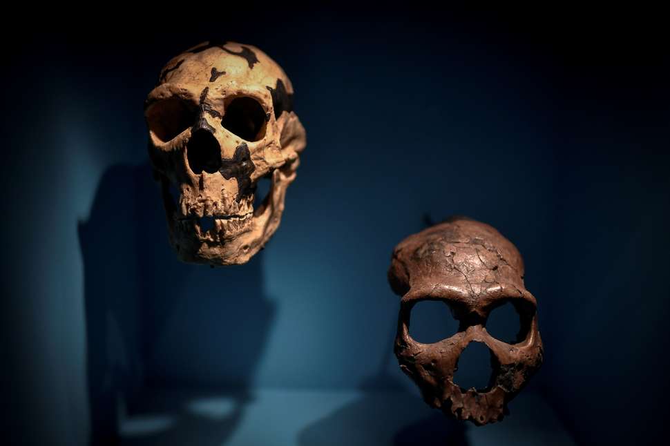
| Skulls are displayed as part of the Neanderthal exhibition at the Musee de l'Homme in Paris on March 26, 2018. |
There are rare cases of ancient brains, such as the 2,600-year-old Heslington Brain, being found "pickled" in certain wet, anoxic environments, but prehistoric brains in the archaeological record are very few.
Scientists therefore lack intact Neanderthal and early Homo sapiens brains to study. But an innovative team has just reconstructed such brains using a technique called computational neuroanatomy. The 3D models they produced, reported in the journal Scientific Reports, are the first of their kind.
"Our attempt to actually reconstruct the brain inside of the fossil crania is completely new to the field," co-author Naomichi Ogihara of Keio University's Department of Mechanical Engineering told Seeker.
Ogihara, co-senior authors Norihiro Sadato and Takeru Akazawa, and their colleagues used virtual casts of four Neanderthal and four early Homo sapiens skull fossils to reconstruct the size of their brains. The Neanderthals lived in what are now Israel, France, and Gibraltar. The early Homo sapiens came from Israel and the Czech Republic.
The authors then used MRI data from the brains of 1,185 living humans to model the average human brain. They also considered non-human primate brains and the skull of a Cro-Magnon individual who lived 32,000 years ago.
The resulting computer model was then deformed to match the shape of the Neanderthal and early Homo sapiens skull casts. This allowed the researchers to predict what the brains of these humans might have looked like, and how individual brain regions could have differed between the two groups.
It should be noted that many researchers believe Neanderthals were members of our species. Ogihara told Seeker there is ample evidence "showing that Neanderthals and Homo sapiens interbred. We believe so, too."
As a result, the majority of people alive today retain Neanderthal DNA. These include people whose heritage is North African, as well as people with Eurasian ancestry.
It is even possible that the early modern humans included in the study were related to Neanderthals.
“We certainly cannot deny the possibility that the specimens we used already interbred with Neanderthals,” Ogihara told Seeker.
But, he added, “there is no obvious reason to think that way, so we basically assumed that the individuals used in the present study had not interbred with Neanderthals.”
The computer models confirmed prior findings that Neanderthal brains were larger than those of anatomically modern humans. The researchers, however, do not believe that bigger is always better when it comes to brains.
The international team concluded that Neanderthals and early modern humans possessed significantly different brain morphologies, including the latter having a larger cerebellum. Since this part of the brain is associated with language comprehension and production, working memory, and cognitive flexibility, the researchers believe that early modern humans were superior to Neanderthals in terms of these abilities.
"We are not saying that Neanderthals were incapable of processing languages," Ogihara said. "We think they could communicate verbally, but their social ability using languages was probably limited because of the brain structural differences."
It is possible that Neanderthals relied more on visual information. They are thought to have been the world's first artists. The earliest known cave art, reported this year, consists of 65,000-year-old paintings found in three Neanderthal caves in Spain.
Ogihara and his colleagues determined that Neanderthals had a larger occipital lobe than did early modern humans.
"The occipital lobe is the visual processing center," Ogihara explained. "Neanderthals possibly required the larger occipital lobe to compensate for low light levels in Europe."
Because of this, Neanderthals may have been unable to evolve the cerebellum expansion seen in early modern humans.
Brain comparison studies come with inherent challenges, as even brains within a particular species today are not the same. The brains of male humans, for example, tend to be slightly larger than those of females, but the majority of scientists believe that brain size does not necessarily correlate with intelligence.
A bigger Neanderthal brain does not appear to have been advantageous when early modern humans began to dominate their former territories. While Neanderthals — via their DNA — were absorbed into modern Homo sapiens to a certain extent, their extinction is widely believed to have begun around 40,000 years ago. This period coincides with greater numbers of early modern humans migrating into Eurasia.
Ogihara said that his team's research cannot definitively conclude what led to the disappearance of Neanderthals. But, he said, the study shows innate morphological differences in the brain structure actually existed between Neanderthals and early Homo sapiens, which possibly led to differences in cognitive and social abilities.
"Although the difference could be subtle,” he said, “such a subtle difference may become significant in terms of natural selection."
The jury is still out on Neanderthal brain power. Joao Zilhao of the Catalan Institute for Research and Advanced Studies was a member of the team that reported the Neanderthal cave art.
Zilhao told Seeker that both Neanderthals and early Homo sapiens must have possessed the cognitive hardware required for advanced symbolic behavior, such as cave art and body ornamentation.
“The fact that we find the capability in both Neandertals and early modern humans implies that said capability existed in the common ancestor around 500,000 years ago,” he said. “Ergo, I would think it entirely logical to consider that the null hypothesis is the co-evolution of brain, language, and symbolic thinking, and that the fundamentals of human cognition as we know it were in place ever since we see people with big brains in the fossil record — i.e., since at least 1.5 million years ago."
Read more at Seeker
Can We Stop a ‘Mass Extinction’ of Human Languages?
![]() |
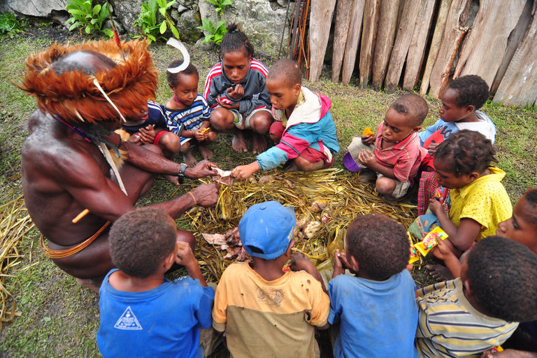
| Tribal people of Papua prepare a feast during Bakar Batu party on February 23, 2015 in Wamena in Papua, Indonesia. |
The rate of language loss has reached such a breakneck pace that some scholars predict we’ll lose 90 percent of the world’s languages in the next century, akin to a linguistic mass extinction event.
It’s not an accident that linguists have borrowed terms from biology to classify languages as vulnerable, endangered, or extinct. As many ethnobiologists and conservationists have come to understand, nature and culture are both products of evolution, and many of the same forces that threaten biological diversity also endanger linguistic diversity.
Jonathan Loh is an honorary research fellow at the University of Kent and the co-author, with Dave Harmon, of a 2014 report for the World Wildlife Fund called “Biocultural Diversity: Threatened Species, Threatened Languages.” Loh and Harmon define biocultural diversity as the sum of evolutionary processes that have produced distinctive species of plants and animals, as well as distinctive cultures and languages. Thanks to shifts in human activity, all are under threat.
The concept that languages evolve in similar ways to biological species isn’t new, Loh told Seeker.
"The formation of different languages and of distinct species, and the proofs that both have been developed through a gradual process, are curiously parallel," Charles Darwin wrote in The Descent of Man. "Dominant languages and dialects spread widely, and lead to the gradual extinction of other tongues. A language, like a species, when once extinct, never, as Sir C. Lyell remarks, reappears."
The reason Darwin was so knowledgeable about the evolution of language, Loh told Seeker, was that a century before Darwin and others were arguing that all species evolved from common ancestors, linguists like William Jones were doing the same thing with language. Jones, an 18th-century British judge in India, spoke more than a dozen languages and took an interest in Ancient Sanskrit, which he discovered had striking similarities to Greek and Latin.
“Which completely blew his mind, because he could think of no reason why there should be,” said Loh.
The answer, Jones decided, was that they must have branched off from some even more ancient tongue, which he called Proto-Indo-European. Jones and others created the first “family tree” of all the languages that diverged and re-converged from that original language — last spoken an estimated 9,000 years ago — including seemingly unrelated languages like Russian, Hindi, Spanish, Swedish, and English.
With the discovery of DNA, biologists began to understand how life on Earth, which began as single-celled organisms 3.9 billion years ago, evolved into the stunning diversity of species on the planet today. Around 540 million years ago, for example, favorable climate and atmospheric conditions led to the Cambrian Explosion, where scientists believe the genetic components came together to jump-start the evolution of multicellular life.
Some time after Homo sapiens were on the scene 200,000 years ago, explained Loh, there was a second explosion — a cultural explosion. And the trigger was the development of language. We don’t know exactly when and where human language first appeared, but language, like DNA, was the vehicle by which information could be passed from one generation to another.
This is where biological evolution and cultural evolution show their fascinating similarities. In natural selection, the gene is the basic currency. If a gene inherited from two parents offers a competitive advantage, it’s more likely to be passed on to the next generation. Biological diversity is powered by constant genetic mutations, which, if advantageous, can branch off into new species.
The evolution of culture, according to influential thinkers like Richard Dawkins and Daniel Dennett, has its own currency: the meme. A meme is unit of a cultural knowledge — like a song, story, recipe, art, or style of dress — that can be passed along primarily through the use of language. Memes, like genes, mutate as they pass from one brain to the next, if these mutated memes gain traction, they may evolve into new cultures and languages.
Interestingly, the regions of the world with the greatest biological diversity are also the ones with the most languages. In general, language diversity follows Rapaport’s Rule, which states that species density is highest at the equator and thins out as you move north and south toward the poles. And there are also distinct “hotspots” of biocultural diversity across the Amazon Basin, Central Africa, and the Indonesia/Malaysia region, home to the undisputed champion of linguistic diversity: New Guinea.
Of the 7,000 languages in the world, 1,000 of them are spoken exclusively in New Guinea. With a population of less than 12 million, that means that 14 percent of the world’s languages are spoken by 0.14 percent of global population.
One of the theories explaining why linguistic diversity blossoms in the tropics is that lots of rivers and mountains divide the landscape, isolating small pockets of people. As Darwin found on the Galapagos Islands, geographic isolation allows for distinct traits to evolve out of the same species. That may help explain the language diversity in New Guinea, said Loh, which is carved up by rivers and mountains, and where tribes are not only isolated, but often hostile to outsiders.
The chief difference between biological and linguistic evolution is the speed of change.
“Biological evolution takes place over millions of years. Languages and cultures evolve incredibly fast by comparison,” said Loh. “If you go back in English to Chaucer, who died only 600 years ago, it’s really hard to read and understand his English. Within 25 generations, that ability to understand has gone, because the language has changed so much.”
Because the rate of change is so fast, languages can also go extinct much faster than biological species. An estimated six percent of global human languages have gone extinct since 1970, for example, while only one percent each of mammal, bird, and amphibian species have disappeared in that same time span.
The main driver of language endangerment and extinction is a process called language shift, when speakers switch from a native, typically indigenous tongue to the dominant national language. John Sullivant, a language data curator with the Archive of the Indigenous Languages of Latin America, told Seeker that language shift happens for a variety of reasons, but is largely driven by the level of contact with the national culture and the marginalization of indigenous communities.
“In Mexico,” Sullivant said, “nearly every indigenous group is in very close contact with Spanish. And that large amount of contact and the ability to move outside of the community — or having to move outside of the community for various reasons — makes the transmission of the language from parents to children that much more precarious.”
It’s clear that economic forces threaten both biological diversity and linguistic diversity. Members of economically marginalized indigenous communities often migrate to bigger cities or even other countries to support their families, shifting to the dominant language for work. Similarly, the globalization of manufacturing increases the plundering of natural resources, which drives habitat loss, one of the main ways that endangered species go extinct.
What’s doubly troubling is that when a language dies out, so does a wealth of knowledge about native plants and animals, exactly the type of information that conservationists need to protect critical species. Some conservation biologists estimate that indigenous communities, which cluster in regions with the greatest natural biodiversity, are the stewards of 99 percent of the world’s genetic diversity.
Richard Stepp is an ethnobiologist at the University of Florida who has conducted fieldwork among indigenous Mayan communities in Mexico, Belize, and Guatemala.
“In some of these cultures, the single largest category of nouns are plant names. They may have thousands of plant names,” Stepp told Seeker. “So the biodiversity is intimately linked to the language.”
Languages, like species, deserve to be preserved for their own sake, but there are also more utilitarian reasons to want to preserve the knowledge encoded in indigenous languages. For example, only a fraction of the world’s plants have been exhaustively studied for their medicinal properties, but it’s very likely that indigenous cultures have cumutively tested just about everything.
“For a lot of these cultures, their primary healthcare is what they find growing around their house,” said Stepp. “In order to know what’s on the shelf of that living pharmacy, you have to have the language.”
Loh believes that more linguists working with indigenous communities need to receive basic training in biology and botany so that they can capture the depth of the scientific knowledge encoded in endangered languages before they disappear. Stepp said that he brings along specialists for that very reason.
“I’ve seen instances where 5-year-old kids know more plants than adult Westerners. They can easily name 150 plant species,” Stepp said. “This knowledge is gained at a really early age, and not only allows them to survive, but to live a very rich life through the knowledge of food plants.”
Read more at Seeker
Apr 26, 2018
Magma ocean may be responsible for the moon's early magnetic field
Scientist Aaron Scheinberg of Princeton, with Krista Soderlund from the University of Texas Institute for Geophysics, and Linda Elkins-Tanton of Arizona State University, set out to determine what may have powered this early lunar magnetic field. Their results and a new model for how this may have happened, have been recently published in Earth and Planetary Science Letters.
A new model
Earth's magnetic field protects our planet by deflecting most of the solar wind, whose charged particles would otherwise strip away the ozone layer that protects the Earth from harmful ultraviolet radiation.
While Earth's magnetic field is generated by the motions of its convecting liquid metal outer core, known as the dynamo, the Moon's core is too small to have produced a magnetic field of that magnitude.
So, the research team proposed a new model for how the magnetic field could have reached Earth-like levels. In this scenario, the dynamo is powered not by the Moon's small metal core, but by a heavy layer of molten (liquid) rock that sits on top of it.
In this proposed model, the bottom-most layer of the Moon's mantle melts to form a metal-rich "basal magma ocean" that sits on top of the Moon's metal core. Convection in this layer then drives the dynamo, creating a magnetic field.
"The idea of a basal magma ocean dynamo had been proposed for the early Earth's magnetic field, and we realized that this mechanism may also be important for the Moon," says co-author Soderlund.
Soderlund further explains that a partially molten layer is thought to still exist at the base of the lunar mantle today. "A strong magnetic field is easier to achieve at the Moon's surface if the dynamo operated in the mantle rather than in the core," she says, "because magnetic field strength decreases rapidly the farther away it is from the dynamo region."
In simulations of the core dynamo of the Moon conducted by the team, they kept finding that the lower layer of the Moon's mantle was overheating and melting. Initially, they tried to focus on cases without melting that were easier to model, but eventually considered that the melting process was the key to their new model.
"Once we started thinking of that melting as a feature, instead of a bug," says Scheinberg, "the pieces started fitting together and we wondered if the melting that we saw in the models could produce a metal-rich magma ocean to power the strong early field."
A later weak magnetic field
Further along in the evolution of the Moon (around 3.56 billion years ago), there is also evidence that the strong magnetic field that existed around the Moon eventually became a weak magnetic field, one that continued until relatively recently. The team's new model may also help explain this phenomenon as well.
"Our model provides an elegant potential solution," says Scheinberg. "As the Moon cooled, the magma ocean would have solidified, while the core dynamo would have continued to create the later weak field."
"We're excited by this result because it explains fundamental observations about the Moon -- its early, strong magnetic field and its subsequent weakening and then disappearance -- using first-order processes already supported by other observations," adds co-author Elkins-Tanton.
Beyond providing a new model to build from, this research may also provide a better understanding of planetary magnetic field generation elsewhere in our solar system and beyond.
Read more at Science Daily
Molecular evolution: How the building blocks of life may form in space
"You just need the right combination of ingredients," author Michael Huels said. "These molecules can combine, they can chemically react, under the right conditions, to form larger molecules which then give rise to the bigger biomolecules we see in cells like components of proteins, RNA or DNA, or phospholipids."
The right conditions, in space, include ionizing radiation. In space, molecules are exposed to UV rays and high-energy radiation including X-rays, gamma rays, stellar and solar wind particles and cosmic rays. They are also exposed to low-energy electrons, or LEEs, produced as a secondary product of the collision between radiation and matter. The authors examined LEEs for a more nuanced understanding of how complex molecules might form.
In their paper, in the Journal of Chemical Physics, from AIP Publishing, the authors exposed multilayer ice composed of carbon dioxide, methane and ammonia to LEEs and then used a type of mass spectrometry called temperature programmed desorption (TPD) to characterize the molecules created by LEEs.
In 2017, using a similar method, these researchers were able to create ethanol, a nonessential molecule, from only two ingredients: methane and oxygen. But these are simple molecules, not nearly as complex as the larger molecules that are the stuff of life. This new experiment has yielded a molecule that is more complex, and is essential for terrestrial life: glycine.
Glycine is an amino acid, made of hydrogen, carbon, nitrogen and oxygen. Showing that LEEs can convert simple molecules into more complex forms illustrates how life's building blocks could have formed in space and then arrived on Earth from material delivered via comet or meteorite impact.
In their experiment, for each 260 electrons of exposure, one molecule of glycine was formed. Seeking to know how realistic this rate of formation was in space, not just in the laboratory, the researchers extrapolated out to determine the probability that a carbon dioxide molecule would encounter both a methane molecule and ammonia molecule and how much radiation they, together, might encounter.
Read more at Science Daily
Archaeologists on ancient horse find in Nile River Valley
The research findings are published in Antiquity. The Tombos horse was discovered in 2011, and members of the Purdue team -- professor Michele Buzon and alumna Sarah Schrader -- played a part in the excavation and analysis. The horse is dated to the Third Intermediate Period, 1050-728 B.C.E., and it was found more than 5 feet underground in a tomb. The horse, with some chestnut-colored fur remaining, had been buried in a funeral position with a burial shroud.
"It was clear that the horse was an intentional burial, which was super fascinating," said Buzon, a professor of anthropology. "Remnants of fabric on the hooves indicate the presence of a burial shroud. Changes on the bones and iron pieces of a bridle suggest that the horse may have pulled a chariot. We hadn't found anything like this in our previous excavations at Tombos. Animal remains are very rare at the site."
Buzon, a bioarchaeologist, has worked with Stuart Tyson Smith, anthropology professor at the University of California, Santa Barbara, for 18 years at this site in modern-day Sudan, and both are principal investigators on the project. Buzon uses health and cultural evidence from more than 3,000-year-old burial sites to understand the lives of Nubians and Egyptians during the New Kingdom Empire. This is when Egyptians colonized the area in about 1500 B.C. to gain access to trade routes on the Nile River. Over the years, hundreds of artifacts, including pottery, tools, carvings and dishes were unearthed at this burial site for about 200 individuals.
"Finding the horse was unexpected," Schrader said. "Initially, we weren't sure if it was modern or not. But as we slowly uncovered the remains, we began to find artifacts associated with the horse, such as the scarab, the shroud and the iron cheekpiece. At that point, we realized how significant this find was. Of course, we became even more excited when the carbon-14 dates were assessed and confirmed how old the horse was."
Schrader, who graduated from Purdue in 2013 with a doctoral degree in anthropology, is an assistant professor of human osteoarchaeology at Leiden University in The Netherlands. Schrader is lead author on this article, and she helped frame this find within the context of Nubian history.
Once the archaeologists discovered the horse, Sandra Olsen, curator-in-charge at the Biodiversity Institute and Natural History Museum at the University of Kansas and a well-known ancient horse expert, was invited to Purdue to analyze the horse skeleton. Buzon coordinated the analysis between the team, and she established the chronology of the horse via radiocarbon dating.
Read more at Science Daily
Subscribe to:
Posts (Atom)