Showing posts with label Nanoparticles. Show all posts

Apr 23, 2024

Breakthrough rice bran nanoparticles show promise as affordable and targeted anticancer agent

Plant-derived nanoparticles have demonstrated significant anticancer effects. Researchers recently developed rice bran-derived nanoparticles (rbNPs) that efficiently suppressed cell proliferation and induced programmed cell death of only cancer cells. Furthermore, rbNPs successfully suppressed the growth of tumors in mice having aggressive adenocarcinoma in their peritoneal cavity, without any adverse effects. Given their low production costs and high efficacy, rbNPs hold great promise for developing affordable and safe anticancer agents.

Several types of conventional cancer therapies, such as radiotherapy or chemotherapy, destroy healthy cells along with cancer cells. In advanced stages of cancer, tissue loss from treatments can be substantial and even fatal. Cutting-edge cancer therapies that employ nanoparticles can specifically target cancer cells, sparing healthy tissue. Recent studies have demonstrated that plant-derived nanoparticles (pdNPs) that have therapeutic effects can be an effective alternative to traditional cancer treatments. However, no pdNPs have been approved as anticancer therapeutic agents till date.

Rice bran is a byproduct generated during rice refining process that has limited utility and low commercial value. However, it contains several compounds with anticancer properties, such as γ-oryzanol and γ-tocotrienol. To explore these therapeutic properties of rice bran, a team of researchers led by Professor Makiya Nishikawa from Tokyo University of Science (TUS) in Japan developed nanoparticles from rice bran and tested their effectiveness in mice models. Their study, published in Volume 22 of Journal of Nanobiotechnology on 16 March 2024, was co-authored by Dr. Daisuke Sasaki, Ms. Hinako Suzuki, Associate Professor Kosuke Kusamori, and Assistant Professor Shoko Itakura from TUS.

"In recent years, an increasing number of new drug modalities are being developed. At the same time, development costs associated with novel therapies have increased dramatically, contributing to the burden of medical expenses. To address this issue, we used rice bran, an industrial waste with anticancer properties, to develop nanoparticles," explains Prof. Nishikawa.

The study evaluated the anticancer effects of rice bran-derived nanoparticles (rbNPs), which were obtained by processing and purifying a suspension of Koshihikari rice bran in water. When a cancer cell line named colon26 was treated with rbNPs, cell division was arrested and programmed cell death was induced, indicating strong anticancer effects of the nanoparticles. The observed anticancer activity of rbNPs can be attributed to γ-tocotrienol and γ-oryzanol, that are easily taken up by cancer cells resulting in cell cycle arrest and programmed cell death. Additionally, rbNPs reduced the expression of proteins, such as β-catenin (a protein associated with Wnt signaling pathway involved in cell proliferation) and cyclin D1, which are known to promote cancer recurrence and metastases. Moreover, the rbNPs reduced the expression of β-catenin only in colon26 cells without affecting the non-cancerous cells.

"A key concern in the context of pdNPs is their low pharmacological activity compared to pharmaceutical drugs. However, rbNPs exhibited higher anticancer activity than DOXIL®, a liposomal pharmaceutical formulation of doxorubicin. Additionally, doxorubicin is cytotoxic to both cancer cells and non-cancerous cells, whereas rbNPs are specifically cytotoxic to cancer cells, suggesting that rbNPs are safer than doxorubicin," highlights Prof.Nishikawa.

To confirm the anticancer properties of rbNPs in the living body, the researchers injected rbNPs into mice having aggressive adenocarcinoma in their peritoneal cavity (enclosed by the diaphragm, abdominal muscles, and pelvis and houses organs like intestines, liver, and kidneys). They observed significant suppression of tumor growth with no adverse effects on the mice. Additionally, the rbNPs significantly inhibited metastatic growth of murine melanoma B16-BL6 cells in a lung metastasis mouse model.

Rice bran has several attributes that make it an excellent source of therapeutic pdNPs. Firstly, it is economic as compared to many other sources of pdNPs. Nearly 40% of the rice bran is discarded in Japan, providing a readily available source of raw material. Secondly, the preparation efficiency of rbNPs is higher than that of previously reported pdNPs. Besides being practical and safe as an anticancer therapeutic, the physicochemical properties of rbNPs are very stable. However, a few parameters, such as establishment of separation technologies at the pharmaceutical level, assessing production process control parameters, and evaluation of efficacy and safety in human cancer cell lines and xenograft animal models, must be investigated prior to clinical trials in humans.

In conclusion, rice bran, an agricultural waste product, is a source of therapeutic pdNPs that are affordable, effective, and safe, and has the potential to revolutionize cancer treatment in the future.

Read more at Science Daily

Feb 19, 2023

How a record-breaking copper catalyst converts CO2 into liquid fuels

Since the 1970s, scientists have known that copper has a special ability to transform carbon dioxide into valuable chemicals and fuels. But for many years, scientists have struggled to understand how this common metal works as an electrocatalyst, a mechanism that uses energy from electrons to chemically transform molecules into different products.

Now, a research team led by Lawrence Berkeley National Laboratory (Berkeley Lab) has gained new insight by capturing real-time movies of copper nanoparticles (copper particles engineered at the scale of a billionth of a meter) as they convert CO2 and water into renewable fuels and chemicals: ethylene, ethanol, and propanol, among others. The work was reported in the journal Nature last week.

"This is very exciting. After decades of work, we're finally able to show -- with undeniable proof -- how copper electrocatalysts excel in CO2 reduction," said Peidong Yang, a senior faculty scientist in Berkeley Lab's Materials Sciences and Chemical Sciences Divisions who led the study. Yang is also a professor of chemistry and materials science and engineering at UC Berkeley. "Knowing how copper is such an excellent electrocatalyst brings us steps closer to turning CO2 into new, renewable solar fuels through artificial photosynthesis."

The work was made possible by combining a new imaging technique called operando 4D electrochemical liquid-cell STEM (scanning transmission electron microscopy) with a soft X-ray probe to investigate the same sample environment: copper nanoparticles in liquid. First author Yao Yang, a UC Berkeley Miller postdoctoral fellow, conceived the groundbreaking approach under the guidance of Peidong Yang while working toward his Ph.D. in chemistry at Cornell University.

Scientists who study artificial photosynthesis materials and reactions have wanted to combine the power of an electron probe with X-rays, but the two techniques typically can't be performed by the same instrument.

Electron microscopes (such as STEM or TEM) use beams of electrons and excel at characterizing the atomic structure in parts of a material. In recent years, 4D STEM (or "2D raster of 2D diffraction patterns using scanning transmission electron microscopy") instruments, such as those at Berkeley Lab's Molecular Foundry, have pushed the boundaries of electron microscopy even further, enabling scientists to map out atomic or molecular regions in a variety of materials, from hard metallic glass to soft, flexible films.

On the other hand, soft (or lower-energy) X-rays are useful for identifying and tracking chemical reactions in real time in an operando, or real-world, environment.

But now, scientists can have the best of both worlds. At the heart of the new technique is an electrochemical "liquid cell" sample holder with remarkable versatility. A thousand times thinner than a human hair, the device is compatible with both STEM and X-ray instruments.

The electrochemical liquid cell's ultrathin design allows reliable imaging of delicate samples while protecting them from electron beam damage. A special electrode custom-designed by co-author Cheng Wang, a staff scientist at Berkeley Lab's Advanced Light Source, enabled the team to conduct X-ray experiments with the electrochemical liquid cell. Combining the two allows researchers to comprehensively characterize electrochemical reactions in real time and at the nanoscale.

Getting granular


During 4D-STEM experiments, Yao Yang and team used the new electrochemical liquid cell to observe copper nanoparticles (ranging in size from 7 nanometers to 18 nanometers) evolve into active nanograins during CO2 electrolysis -- a process that uses electricity to drive a reaction on the surface of an electrocatalyst.

The experiments revealed a surprise: copper nanoparticles combined into larger metallic copper "nanograins" within seconds of the electrochemical reaction.

To learn more, the team turned to Wang, who pioneered a technique known as "resonant soft X-ray scattering (RSoXS) for soft materials," at the Advanced Light Source more than 10 years ago.

With help from Wang, the research team used the same electrochemical liquid cell, but this time during RSoXS experiments, to determine whether copper nanograins facilitate CO2 reduction. Soft X-rays are ideal for studying how copper electrocatalysts evolve during CO2 reduction, Wang explained. By using RSoXS, researchers can monitor multiple reactions between thousands of nanoparticles in real time, and accurately identify chemical reactants and products.

The RSoXS experiments at the Advanced Light Source -- along with additional evidence gathered at Cornell High Energy Synchrotron Source (CHESS) -- proved that metallic copper nanograins serve as active sites for CO2 reduction. (Metallic copper, also known as copper(0), is a form of the element copper.)

During CO2 electrolysis, the copper nanoparticles change their structure during a process called "electrochemical scrambling." The copper nanoparticles' surface layer of oxide degrades, creating open sites on the copper surface for CO2 molecules to attach, explained Peidong Yang. And as CO2 "docks" or binds to the copper nanograin surface, electrons are then transferred to CO2, causing a reaction that simultaneously produces ethylene, ethanol, and propanol along with other multicarbon products.

"The copper nanograins essentially turn into little chemical manufacturing factories," Yao Yang said.

Further experiments at the Molecular Foundry, the Advanced Light Source, and CHESS revealed that size matters. All of the 7-nanometer copper nanoparticles participated in CO2 reduction, whereas the larger nanoparticles did not. In addition, the team learned that only metallic copper can efficiently reduce CO2 into multicarbon products. The findings have implications for "rationally designing efficient CO2 electrocatalysts," Peidong Yang said.

The new study also validated Peidong Yang's findings from 2017: That the 7-nanometer-sized copper nanoparticles require low inputs of energy to start CO2 reduction. As an electrocatalyst, the 7-nanometer copper nanoparticles required a record-low driving force that is about 300 millivolts less than typical bulk copper electrocatalysts. The best-performing catalysts that produce multicarbon products from CO2 typically operate at high driving force of 1 volt.

The copper nanograins could potentially boost the energy efficiency and productivity of some catalysts designed for artificial photosynthesis, a field of research that aims to produce solar fuels from sunlight, water, and CO2. Currently, researchers within the Department of Energy-funded Liquid Sunlight Alliance (LiSA) plan to use the copper nanograin catalysts in the design of future solar fuel devices.

"The technique's ability to record real-time movies of a chemical process opens up exciting opportunities to study many other electrochemical energy conversion processes. It's a huge breakthrough, and it would not have been possible without Yao and his pioneering work," Peidong Yang said.

Read more at Science Daily

Jul 11, 2022

Nanoparticles can save historic buildings

Many historical buildings were built of limestone, such as Vienna's St. Stephen's Cathedral. Limestone is easy to work with, but does not withstand weathering well. It consists mainly of calcite minerals that are relatively weakly bound to each other, which is why parts of the stone keep crumbling away over the years, often requiring costly restoration and conservation treatments.

However, it is possible to increase the resistance of the stone by treating it with special silicate nanoparticles. The method is already being used, but what exactly happens in the process and which nanoparticles are best suited for this purpose has been unclear until now. A research team from TU Wien and the University of Oslo has now been able to clarify exactly how this artificial hardening process takes place through elaborate experiments at the DESY synchrotron in Hamburg and with microscopic examinations in Vienna. That way, the team could determine which nanoparticles are best suited for this purpose.

An aqueous suspension with nanoparticles

"We use a suspension, a liquid, in which the nanoparticles initially float around freely," says Prof. Markus Valtiner from the Institute of Applied Physics at TU Wien. "When this suspension gets into the rock, then the aqueous part evaporates, the nanoparticles form stable bridges between the minerals and give the rock additional stability."

This method is already used in restoration technology, but until now it was not known exactly what physical processes take place. When the water evaporates, a very special kind of crystallisation occurs: Normally, a crystal is a regular arrangement of individual atoms. However, not only atoms, but also entire nanoparticles can arrange themselves in a regular structure -- this is then referred to as a "colloidal crystal."

The silicate nanoparticles come together to form such colloidal crystals when they dry in the rock and thus jointly create new connections between the individual mineral surfaces. This increases the strength of the natural stone.

Measurements at the large-scale research facility DESY and in Vienna

To observe this crystallisation process in detail, the TU Wien research team used the DESY synchrotron facility in Hamburg. Extremely strong X-rays can be generated there, which can be used to analyse the crystallisation during the drying process.

"This was very important to understand exactly what the strength of the bonds that form depends on," says Joanna Dziadkowiec (University of Oslo and TU Wien), the first author of the publication in which the research results have now been presented. "We used nanoparticles of different sizes and concentrations and studied the crystallisation process with X-ray analyses." It was shown that the size of the particles is decisive for optimal strength gain.

To this end, the TU Vienna also measured the adhesive force created by the colloidal crystals. For this purpose, a special interference microscope was used, which is perfectly suited for measuring tiny forces between two surfaces.

Read more at Science Daily

Apr 18, 2022

Extract from a common kitchen spice could be key to greener, more efficient fuel cells

Turmeric, a spice found in most kitchens, has an extract that could lead to safer, more efficient fuel cells.

Researchers at the Clemson Nanomaterials Institute (CNI) and their collaborators from the Sri Sathya Sai Institute of Higher Learning (SSSIHL) in India discovered a novel way to combine curcumin -- the substance in turmeric -- and gold nanoparticles to create an electrode that requires 100 times less energy to efficiently convert ethanol into electricity.

While the research team must do more testing, the discovery brings replacing hydrogen as a fuel cell feedstock one step closer.

"Of all the catalysts for alcohol oxidation in alkaline medium, the one we prepared is the best so far," said Apparao Rao, CNI's founding director and the R. A. Bowen Professor of Physics in the College of Science's.

Fuel cells generate electricity through a chemical reaction instead of combustion. They are used to power vehicles, buildings, portable electronic devices and backup power systems.

Hydrogen fuel cells are highly efficient and do not produce greenhouse gases. While hydrogen is the most common chemical element in the universe, it must be derived from substances such as natural gas and fossil fuels because it occurs naturally on Earth only in compound form with other elements in liquids, gases or solids. The necessary extraction adds to hydrogen fuel cells' cost and environmental impact.

In addition, hydrogen used in fuel cells is a compressed gas, creating challenges for storage and transportation. Ethanol, an alcohol made from corn or other agricultural-based feeds, is safer and easier to transport than hydrogen because it is a liquid.

"To make it a commercial product where we can fill our tanks with ethanol, the electrodes have to be highly efficient," said Lakshman Ventrapragada, a former student of Rao's who worked as a research assistant at the CNI and is an alumnus of SSSIHL. "At the same time, we don't want very expensive electrodes or synthetic polymeric substrates that are not eco-friendly because that defeats the whole purpose. We wanted to look at something green for the fuel cell generation process and making the fuel cell itself."

The researchers focused on the fuel cell's anode, where the ethanol or other feed source is oxidized.

Fuel cells widely use platinum as a catalyst. But platinum suffers from poisoning because of reaction intermediates such as carbon monoxide, Ventrapragada said. It is also costly.

The researchers used gold as a catalyst. Instead of using conducting polymers, metal-organic frameworks, or other complex materials to deposit the gold on the surface of the electrode, the researchers used curcumin because of its structural uniqueness. Curcumin is used to decorate the gold nanoparticles to stabilize them, forming a porous network around the nanoparticles. Researchers deposited the curcumin gold nanoparticle on the surface of the electrode at a 100 times lower electric current than in previous studies.

Without the curcumin coating, the gold nanoparticles agglomerate, cutting down on the surface area exposed to the chemical reaction, Ventrapragada said.

"Without this curcumin coating, the performance is poor," Rao said. "We need this coating to stabilize and create a porous environment around the nanoparticles, and then they do a super job with alcohol oxidation.

"There's a big push in the industry for alcohol oxidation. This discovery is an excellent enabler for that. The next step is to scale the process up and work with an industrial collaborator who can actually make the fuel cells and build stacks of fuel cells for the real application," he continued.

But the research could have broader implications than improved fuel cells. The electrode's unique properties could lend itself to future applications in sensors, supercapacitors and more, Ventrapragada said.

In collaboration with the SSSIHL research team, Rao's team is testing the electrode as a sensor that could help identify changes in the level of dopamine. Dopamine has been implicated in disorders such as Parkinson's disease and attention deficit hyperactivity disorder. When members of the research team tested urine samples obtained from healthy volunteers, they could measure dopamine to the approved clinical range with this electrode using a cost-effective method compared to standard ones used today, Rao said.

Read more at Science Daily

Jan 22, 2022

Novel nanoantibiotics kill bacteria without harming healthy cells

The Centers for Disease Control and Prevention estimates that more than 2.8 million Americans experience antibiotic-resistant infections each year; more than 35,000 die from those infections.

To address this critical and worldwide public health issue, a team of researchers led by Hongjun (Henry) Liang, Ph.D., from the Texas Tech University Health Sciences Center (TTUHSC) Department of Cell Physiology and Molecular Biophysics, recently investigated whether or not a series of novel nanoparticles can kill some of the pathogens that lead to human infection without affecting healthy cells.

The study, "Hydrophilic Nanoparticles that Kill Bacteria while Sparing Mammalian Cells Reveal the Antibiotic Role of Nanostructures," was published Jan. 11 by Nature Communications. Other study members of the Liang team, all from TTUHSC, included Yunjiang Jiang, Ph.D., Wan Zheng, Ph.D., Keith Tran, Elizabeth Kamilar, Jitender Bariwal, Ph.D., and Hairong Ma, Ph.D.

Past research has shown that hydrophobicity (a molecule's ability to repel water) and hydrophilicity (a molecule's ability to attract and dissolve in water) affects cells; the more hydrophobic a substance is, the more adverse the reaction it will cause. However, Liang said, there is no quantitative standard for how much hydrophobicity is acceptable.

"Basically, you can kill bacteria when you increase hydrophobicity," Liang said. "But it will also kill healthy cells, and we don't want that."

For their study, the Liang team used novel hydrophilic nanoparticles known as nanoantibiotics that were developed by Liang's laboratory. Structurally speaking, these novel nanoantibiotics resemble tiny hairy spheres, each composed of many hydrophilic polymer brushes grafted onto silica nanoparticles of different sizes.

These synthetic compounds, which Liang's lab produces, are designed to kill bacteria via membrane disruptions like antimicrobial peptides do, but through a different mode of membrane remodeling that damages bacterial membranes and not mammalian cells. Antimicrobial peptides are a diverse class of amphipathic molecules (partially hydrophilic-partially hydrophobic), which occur naturally and serve as the first line of defense for all multicellular organisms. The direct use of antimicrobial peptides as antibiotics is limited by their stability and toxicity.

There have been other studies in which researchers grafted amphipathic molecules onto nanoparticles, and they too kill bacteria. However, Liang said the primary issue in using amphipathic molecules is that it becomes very difficult to strike the right balance between their hydrophobicity and hydrophilicity so that the toxicity of these molecules to our own cells is significantly reduced.

"In our case, we remove that uncertainty from the equation because we started with a hydrophilic polymer," Liang pointed out. "The cytotoxicity of hydrophobic moieties is not a concern anymore. Those hydrophilic polymers by themselves, or the silica nanoparticles alone don't kill bacteria; they have to be grafted onto the nanostructure to be able to kill bacteria. And so, this is the first important discovery."

The Liang team also discovered that the degree of antibiotic activity is affected by the size of the hairy spheres, which according to Liang is the second important discovery of this research. Those measuring 50 nanometers and below appear to be much more active than those whose size exceeds 50 nanometers. Liang said those measuring approximately 10 nanometers appear to be the most active. (Using synchrotron small angle x-ray scattering and other methods, the Liang team is able to interpret the molecular mechanism of the size-dependent antibiotic activity.)

These discoveries are important because using nanoantibiotics to kill bacteria evades all known mechanisms of bacterial resistance unless bacteria completely revamp their pathways for making cell membranes, which Liang said is unlikely.

Read more at Science Daily

Jan 4, 2022

First time genome editing made possible on cells lining blood vessel walls

The lab of Youyang Zhao, PhD, from Stanley Manne Children's Research Institute at Ann & Robert H. Lurie Children's Hospital of Chicago developed a unique nanoparticle to deliver genome editing technology, including CRISPR/Cas9, to endothelial cells, which are cells that line blood vessel walls. This is the first time that vascular endothelial cells could be reached for genome editing, since the usual way to deliver CRISPR/Cas9 -- through a virus -- does not work for this cell type. Findings were published in the journal Cell Reports.

"The nanoparticle we developed is a powerful new delivery system for genome editing in vascular endothelial cells, and could be used to treat many diseases, including acute respiratory distress syndrome from severe COVID-19," said senior author Dr. Zhao from Lurie Children's. "With this nanoparticle we can introduce genes to inhibit vascular injury and/or promote vascular repair, correct gene mutations and turn genes on or off to restore normal function. It also allows us to edit multiple genes at the same time. This is an important advance for treating any disease caused by endothelial dysfunction."

Endothelial dysfunction is at the root of many diseases, such as coronary artery disease, stroke, bronchopulmonary dysplasia and pulmonary artery hypertension. Dr. Zhao explained that genome editing in endothelial cells could even treat cancers by cutting off the blood supply to the tumor or blocking cancer metastasis.

At this stage, Dr. Zhao and colleagues achieved excellent results in a mouse model. The nanoparticle carrying CRISPR/Cas9 plasmid DNA was introduced via a one-time IV injection and required a few days to be effective. Preclinical testing will be necessary before clinical trials can begin.

"Our nanoparticle delivery system for genome editing and transgene expression also is a huge advance for cardiovascular research," added Dr. Zhao.

From Science Daily

Dec 29, 2021

Researchers develop structural blueprint of nanoparticles to target white blood cells responsible for acute lung inflammation

The COVID-19 pandemic highlighted the devastating impact of acute lung inflammation (ALI), which is part of the acute respiratory distress syndrome (ARDS) that is the dominant cause of death in COVID-19. A potential new route to the diagnosis and treatment of ARDS comes from studying how neutrophils -- the white blood cells responsible for detecting and eliminating harmful particles in the body -- differentiate what materials to uptake by the material's surface structure, and favor uptake of particles that exhibit "protein clumping," according to new research from the Perelman School of Medicine at the University of Pennsylvania. The findings are published in Nature Nanotechnology.

Researchers investigated how neutrophils are able to differentiate between bacteria to be destroyed and other compounds in the bloodstream, such as cholesterol particles. They tested a library consisting of 23 different protein-based nanoparticles in mice with ALI which revealed a set of "rules" that predict uptake by neutrophils. Neutrophils don't take up symmetrical, rigid particles, such as viruses, but they do take up particles that exhibited "protein clumping," which the researchers call nanoparticles with agglutinated protein (NAPs).

"We want to utilize the existing function of neutrophils that identifies and eliminates invaders to inform how to design a 'Trojan horse' nanoparticle that overactive neutrophils will intake and deliver treatment to alleviate ALI and ARDS," said study lead author Jacob Myerson, PhD, a postdoctoral research fellow in the Department of Systems Pharmacology and Translational Therapeutics. "In order to build this 'Trojan horse' delivery system, though, we had to determine how neutrophils identify which particles in the blood to take up."

ALI and ARDS are life-threatening forms of respiratory failure with high morbidity and mortality rates. Prior to COVID-19, there were 190,000 annual cases of ARDS in the U.S. and 75,000 deaths, with the ARDS being caused by pneumonia, sepsis, and trauma. However, COVID has increased ARDS cases into the millions. When ALI or ARDS occurs, the lung's air sacs recruit neutrophils to the lungs in order to eliminate circulating microbes. This process causes neutrophils to release compounds that further aggravate lung injury and damage the air sacs, so patients develop low blood oxygen levels. Unfortunately, despite the severity of ALI/ARDS, there is no effective drug to control it, and treatment currently focuses on supporting patients while the lungs naturally, but slowly, heal.

To address ARDS and other medical problems, researchers at Penn and elsewhere have been using nanoparticles to concentrate drugs in injured or diseased organs. Such nanoparticles are also being used for gene therapy and immunotherapy.

The researchers note that while the development of viable therapies for ALI/ARDS using nanoparticles to deliver treatments via neutrophils are a long way off, this research represents a significant step in understanding the condition and function of the immune system.

"Now that we have determined that neutrophils patrol for nanoparticles with agglutinated protein, our next step is to understand how and why other microbes, like viruses, which are rigid and symmetrical, evolved to evade neutrophils," said senior author Jacob Brenner, MD, PhD, an associate professor of Pulmonary Medicine in the Division of Pulmonary, Allergy, and Critical Care. "With this knowledge, we can continue to utilize this unique combination of material science and engineering, to create disease-specific therapies that target more advanced and complicated pathologies."

Read more at Science Daily

Dec 13, 2021

'Supermeres' may carry clues to cancer, Alzheimer's disease and COVID-19

Researchers at Vanderbilt University Medical Center have discovered a nanoparticle released from cells, called a "supermere," which contains enzymes, proteins and RNA associated with multiple cancers, cardiovascular disease, Alzheimer's disease and even COVID-19.

The discovery, reported in Nature Cell Biology, is a significant advance in understanding the role extracellular vesicles and nanoparticles play in shuttling important chemical "messages" between cells, both in health and disease.

"We've identified a number of biomarkers and therapeutic targets in cancer and potentially in a number of other disease states that are cargo in these supermeres," said the paper's senior author, Robert Coffey, MD. "What is left to do now is to figure out how these things get released."

Coffey, the Ingram Professor of Cancer Research and professor of Medicine and Cell & Developmental Biology, is internationally known for his studies of colorectal cancer. His team is currently exploring whether the detection and targeting of cancer-specific nanoparticles in the bloodstream could lead to earlier diagnoses and more effective treatment.

In 2019 Dennis Jeppesen, PhD, a former research fellow in Coffey's lab who is now a research instructor in Medicine, used advanced techniques to isolate and analyze small membrane-enclosed extracellular vesicles called "exosomes."

That year, using high-speed ultracentrifugation, another of Coffey's colleagues, Qin Zhang, PhD, research assistant professor of Medicine, devised a simple method to isolate a nanoparticle called an "exomere" that lacks a surface coat.

In the current study, Zhang took the "supernatant," or fluid that remains after the exomeres have been spun into a "pellet," and spun the fluid faster and longer.

The result was a pellet of nanoparticles isolated from the supernatant of the exomere spin -- which the researchers named supermeres. "They're also super-interesting," Coffey quipped, "because they contain many cargo previously thought to be in exosomes."

For one thing, supermeres carry most of the extracellular RNA released by cells and which is found in the bloodstream. Among other functional properties, cancer-derived supermeres can "transfer" drug resistance to tumor cells, perhaps via the RNA cargo they deliver, the researchers reported.

Supermeres are important carriers of TGFBI, a protein that in established tumors promotes tumor progression. TGFBI thus may be a useful marker in liquid biopsies for patients with colorectal cancer, the researchers noted.

They also carry ACE2, a cell-surface receptor that plays a role in cardiovascular disease and is the target of the COVID-19 virus. This raises the possibility that ACE2 carried by supermeres could serve as a "decoy" to bind the virus and prevent infection.

Another potentially important cargo is APP, the amyloid-beta precursor protein implicated in the development of Alzheimer's disease. Supermeres can cross the blood-brain barrier, suggesting that their analysis could improve early diagnosis or possibly even targeted treatment of the disease.

"The identification of this rich plethora of bioactive molecules … raises interesting questions about the function of supermeres, and heightens interest in the potential of these particles as biomarkers for diseases," researchers at the University of Notre Dame noted in a review published with the paper.

Read more at Science Daily

Dec 4, 2021

Immune system-stimulating nanoparticle could lead to more powerful vaccines

A common strategy to make vaccines more powerful is to deliver them along with an adjuvant -- a compound that stimulates the immune system to produce a stronger response.

Researchers from MIT, the La Jolla Institute for Immunology, and other institutions have now designed a new nanoparticle adjuvant that may be more potent than others now in use. Studies in mice showed that it significantly improved antibody production following vaccination against HIV, diphtheria, and influenza.

"We started looking at this particular formulation and found that it was incredibly potent, better than almost anything else we had tried," says Darrell Irvine, the Underwood-Prescott Professor with appointments in MIT's departments of Biological Engineering and Materials Science and Engineering; an associate director of MIT's Koch Institute for Integrative Cancer Research; and a member of the Ragon Institute of MGH, MIT, and Harvard.

The researchers now hope to incorporate the adjuvant into an HIV vaccine that is currently being tested in clinical trials, in hopes of improving its performance.

Irvine and Shane Crotty, a professor at the Center for Infectious Disease and Vaccine Research at the La Jolla Institute for Immunology, are the senior authors of the study, which appears today in Science Immunology. The lead authors of the paper are Murillo Silva, a former MIT postdoc, and Yu Kato, a staff scientist at the La Jolla Institute.

More powerful vaccines

Although the idea of using adjuvants to boost vaccine effectiveness has been around for decades, there are only a handful of FDA-approved vaccine adjuvants. One is aluminum hydroxide, an aluminum salt that induces inflammation, and another is an oil and water emulsion that is used in flu vaccines. A few years ago, the FDA approved an adjuvant based on saponin, a compound derived from the bark of the Chilean soapbark tree.

Saponin formulated in liposomes is now used as an adjuvant in the shingles vaccine, and saponins are also being used in a cage-like nanoparticle called an immunostimulatory complex (ISCOM) in a Covid-19 vaccine that is currently in clinical trials.

Researchers have shown that saponins promote inflammatory immune responses and stimulate antibody production, but how they do that is unclear. In the new study, the MIT and La Jolla team wanted to figure out how the adjuvant exerts its effects, and to see if they could make it more potent.

They designed a new type of adjuvant that is similar to the ISCOM adjuvant but also incorporates a molecule called MPLA, which is a toll-like receptor agonist. When these molecules bind to toll-like receptors on immune cells, they promote inflammation. The researchers call their new adjuvant SMNP (saponin/MPLA nanoparticles).

"We expected that this could be interesting because saponin and toll-like receptor agonists are both adjuvants that have been studied separately and shown to be very effective," Irvine says.

The researchers tested the adjuvant by injecting it into mice along with a few different antigens, or fragments of viral proteins. These included two HIV antigens, as well as diphtheria and influenza antigens. They compared the adjuvant to several other approved adjuvants and found that the new saponin-based nanoparticle elicited a stronger antibody response than any of the others.

One of the HIV antigens that they used is an HIV envelope protein nanoparticle, which presents many copies of the gp120 antigen that is present on the HIV viral surface. This antigen recently completed initial testing in phase 1 clinical trials. Irvine and Crotty are part of the Consortium for HIV/AIDS Vaccine Development at the Scripps Research Institute, which ran that trial. The researchers now hope to develop a way to manufacture the new adjuvant at large scale so it can be tested along with an HIV envelope trimer in another clinical trial beginning next year. Clinical trials that combine envelope trimers with the traditional vaccine adjuvant aluminum hydroxide are also underway.

"Aluminum hydroxide is safe but not particularly potent, so we hope that (the new adjuvant) would be an interesting alternative to elicit neutralizing antibody responses in people," Irvine says.

Rapid flow

When vaccines are injected into the arm, they travel through lymph vessels to the lymph nodes, where they encounter and activate B cells. The research team found that the new adjuvant speeds up the flow of lymph to the nodes, helping the antigen to get there before it starts to break down. It does this in part by stimulating immune cells called mast cells, which previously were not known to be involved in vaccine responses.

"Getting to the lymph nodes quickly is useful because once you inject the antigen, it starts slowly breaking down. The sooner a B cell can see that antigen, the more likely it's fully intact, so that B cells are targeting the structure as it will be present on the native virus," Irvine says.

Additionally, once the vaccine reaches the lymph nodes, the adjuvant causes a layer of cells called macrophages, which act as a barrier, to die off quickly, making it easier for the antigen to get into the nodes.

Another way that the adjuvant helps boost immune responses is by activating inflammatory cytokines that drive a stronger response. The TLR agonist that the researchers included in the adjuvant is believed to amplify that cytokine response, but the exact mechanism for that is not known yet.

This kind of adjuvant could also be useful for any other kind of subunit vaccine, which consists of fragments of viral proteins or other molecules. In addition to their work on HIV vaccines, the researchers are also working on a potential Covid-19 vaccine, along with J. Christopher Love's lab at the Koch Institute. The new adjuvant also appears to help stimulate T cell activity, which could make it useful as a component of cancer vaccines, which aim to stimulate the body's own T cells to attack tumors.

Read more at Science Daily

Aug 22, 2021

Here comes the Sun: Planetary scientists find evidence of solar-driven change on the Moon

Tiny iron nanoparticles unlike any found naturally on Earth are nearly everywhere on the Moon -- and scientists are trying to understand why. A new study led by Northern Arizona University doctoral candidate Christian J. Tai Udovicic, in collaboration with associate professor Christopher Edwards, both of NAU's Department of Astronomy and Planetary Science, uncovered important clues to help understand the surprisingly active lunar surface. In an article recently published in Geophysical Research Letters, the scientists found that solar radiation could be a more important source of lunar iron nanoparticles than previously thought.

Asteroid impacts and solar radiation affect the Moon in unique ways because it lacks the protective magnetic field and atmosphere that protect us here on Earth. Both asteroids and solar radiation break down lunar rocks and soil, forming iron nanoparticles (some smaller, some larger) that are detectable from instruments on satellites orbiting the Moon. The study used data from National Aeronautics and Space Administration (NASA) and Japan Aerospace Exploration Agency (JAXA) spacecraft to understand how quickly iron nanoparticles form on the Moon over time.

"We have thought for a long time that the solar wind has a small effect on lunar surface evolution, when in fact it may be the most important process producing iron nanoparticles," Tai Udovicic said. "Since iron absorbs a lot of light, very small amounts of these particles can be detected from very far away -- making them a great indicator of change on the Moon."

Surprisingly, the smaller iron nanoparticles seemed to form at a similar rate as radiation damage in samples returned from the Apollo missions to the Moon, a hint that the Sun has a strong influence in their formation.

"When I saw the Apollo sample data and our satellite data side by side for the first time, I was shocked," Tai Udovicic said. "This study shows that the solar radiation could have a much larger influence in active change on the Moon than previously thought, not only darkening its surface, but it might also create small quantities of water usable in future missions."

As NASA prepares to land the first woman and the next man on the surface of the Moon by 2024 as part of the Artemis mission, understanding the solar radiation environment and possible resources on the Moon are critical. In future work recently awarded a NASA Future Investigators in Space Science and Technology (FINESST) grant, Tai Udovicic plans to broaden his targeted study to the entire Moon, but is also eager to take a closer look at mysterious lunar swirls, one of which was recently selected as a landing site for the upcoming Lunar Vertex rover. He also studies lunar temperatures and water ice stability to inform future missions.

Read more at Science Daily

Jan 21, 2020

Addressing global warming with new nanoparticles and sunshine

Harvesting sunlight, researchers of the Center for Integrated Nanostructure Physics, within the Institute for Basic Science (IBS, South Korea) published in Materials Today a new strategy to transform carbon dioxide (CO2) into oxygen (O2) and pure carbon monoxide (CO) without side-products in water. This artificial photosynthesis method could bring new solutions to environmental pollution and global warming.

While, in green plants, photosynthesis fixes CO2 into sugars, the artificial photosynthesis reported in this study can convert CO2 into oxygen and pure CO as output. The latter can then be employed for a broad range of applications in electronics, semiconductor, pharmaceutical, and chemical industries. The key is to find the right high-performance photocatalyst to help the photosynthesis take place by absorbing light, convert CO2, and ensuring an efficient flow of electrons, which is essential for the entire system.

Titanium oxide (TiO2) is a well-known photocatalyst. It has already attracted significant attention in the fields of solar energy conversion and environmental protection due to its high reactivity, low toxicity, chemical stability, and low cost. While conventional TiO2 can absorb only UV light, the IBS research team reported previously two different types of blue-colored TiO2 (or "blue titania") nanoparticles that could absorb visible light thanks to a reduced bandgap of about 2.7 eV. They were made of ordered anatase/disordered rutile (Ao/Rd) TiO2 (called, HYL's blue TiO2-I) (Energy & Environmental Science, 2016), and disordered anatase/ordered rutile (Ad/Ro) TiO2 (called, HYL's blue TiO2-II) (ACS Applied Materials & Interfaces, 2019), where anatase and rutile refer to two crystalline forms of TiO2 and the introduction of irregularities (disorder) in the crystal enhances the absorption of visible and infra-red light.

For the efficient artificial photosynthesis for the conversion of CO2 into oxygen and pure CO, IBS researchers aimed to improve the performance of these nanoparticles by combining blue (Ao/Rd) TiO2 with other semiconductors and metals that can enhance water oxidation to oxygen, in parallel to CO2 reduction into CO only. The research team obtained the best results with hybrid nanoparticles made of blue titania, tungsten trioxide (WO3), and 1% silver (TiO2/WO3-Ag). WO3 was chosen because of the low valence band position with its narrow bandgap of 2.6 eV, high stability, and low cost. Silver was added because it enhances visible light absorption, by creating a collective oscillation of free electrons excited by light, and also gives high CO selectivity. The hybrid nanoparticles showed about 200 times higher performance than nanoparticles made of TiO2 alone and TiO2/WO3 without silver.

Read more at Science Daily

Oct 1, 2018

Eco-friendly nanoparticles for artificial photosynthesis

This is a schematic representation of photocatalytic hydrogen production with InP/ZnS quantum dots in a typical assay.
Researchers at the University of Zurich have developed a nanoparticle type for novel use in artificial photosynthesis by adding zinc sulfide on the surface of indium-based quantum dots. These quantum dots produce clean hydrogen fuel from water and sunlight -- a sustainable source of energy. They introduce new eco-friendly and powerful materials to solar photocatalysis.

Quantum dots are true all-rounders. These material structures, which are only a few nanometers in size, display a similar behavior to that of molecules or atoms, and their form, size and number of electrons can be modulated systematically. This means that their electrical and optical characteristics can be customized for a number of target areas, such as new display technologies, biomedical applications as well as photovoltaics and photocatalysis.

Fuel production using sunlight and water

Another current line of application-oriented research aims to generate hydrogen directly from water and solar light. Hydrogen, a clean and efficient energy source, can be converted into forms of fuel that are used widely, including methanol and gasoline. The most promising types of quantum dots previously used in energy research contain cadmium, which has been banned from many commodities due to its toxicity. The team of Greta Patzke, Professor at the Department of Chemistry of the University of Zurich, and scientists from Southwest Petroleum University in Chengdu and the Chinese Academy of Sciences have now developed a new type of nanomaterials without toxic components for photocatalysis.

Indium-containing core with a thin layer of zinc sulfide

The three-nanometer particles consist of a core of indium phosphide with a very thin surrounding layer of zinc sulfide and sulfide ligands. "Compared to the quantum dots that contain cadmium, the new composites are not only environmentally friendly, but also highly efficient when it comes to producing hydrogen from light and water," explains Greta Patzke. Sulfide ligands on the quantum dot surface were found to facilitate the crucial steps involved in light-driven chemical reactions, namely the efficient separation of charge carriers and their rapid transfer to the nanoparticle surface.

Great potential for eco-friendly applications

The newly developed cadmium-free nanomaterials have the potential to serve as a more eco-friendly alternative for a variety of commercial fields. "The water-soluble and biocompatible indium-based quantum dots can in the future also be tested in terms of biomass conversion to hydrogen. Or they could be developed into low-toxic biosensors or non-linear optical materials, for example," adds Greta Patzke. She will continue to focus on the development of catalysts for artificial photosynthesis within the University Research Priority Program "LightChEC." This interdisciplinary research program aims to develop new molecules, materials and processes for the direct storage of solar light energy in chemical bonds.

Read more at Science Daily

Jan 24, 2018

Nanoparticle vaccine offers universal protection against influenza A viruses, study finds

Vaccine
Researchers have developed a universal vaccine to combat influenza A viruses that produces long-lasting immunity in mice and protects them against the limitations of seasonal flu vaccines, according to a study led by Georgia State University.

Influenza, a contagious respiratory illness that infects the nose, throat and lungs, is among the leading causes of death in the United States, according to the Centers for Disease Control and Prevention (CDC). The CDC estimates influenza has resulted in between 12,000 and 56,000 deaths annually in the U.S. since 2010.

Seasonal flu vaccines must be updated each year to match the influenza viruses that are predicted to be most common during the upcoming flu season, but protection doesn't always meet expectations or new viruses emerge and manufacturers incorrectly guess which viruses will end up spreading. In 2009, the H1N1 pandemic caused 200,000 deaths during the first 12 months, and low vaccine effectiveness was also observed during the 2014-15 and 2016-17 flu seasons. A universal flu vaccine that offers broad protection against various viruses is urgently needed and would eliminate the limitations of seasonal flu vaccines.

Seasonal flu vaccines provide protective immunity against influenza viruses by targeting the exterior head of the virus's surface protein, which is hemagglutinin (HA). The influenza virus trains the body to produce antibodies against inactivated virus particles containing the head of this protein, ideally preventing the head from attaching to receptors and stopping infection. However, the head is highly variable and is different for each virus, creating a need for better vaccines. This study uses a new approach and instead targets the inside portion of the HA protein known as the stalk, which is more conservative and offers the opportunity for universal protection.

In this study, the researchers found vaccinating mice with double-layered protein nanoparticles that target the stalk of this protein produces long-lasting immunity and fully protects them against various influenza A viruses. The findings are published in the journal Nature Communications.

"Vaccination is the most effective way to prevent deaths from influenza virus, but the virus changes very fast and you have to receive a new vaccination each year," said Dr. Bao-Zhong Wang, associate professor in the Institute for Biomedical Sciences at Georgia State. "We're trying to develop a new vaccine approach that eliminates the need for vaccination every year. We're developing a universal influenza vaccine. You wouldn't need to change the vaccine type every year because it's universal and can protect against any influenza virus.

"What we wanted to do is to induce responses to this stalk part of the influenza surface glycoprotein, not the head part. This way you're protected against different viruses because all influenza viruses share this stalk domain. However, this stalk domain itself isn't stable, so we used a very special way to make this vaccine construct with the stalk domain and had success. We assembled this stalk domain into a protein nanoparticle as a vaccine. Once inside, the nanoparticle can protect this antigenic protein so it won't be degraded. Our immune cells have a good ability to take in this nanoparticle, so this nanoparticle is much, much better than a soluble protein to induce immune responses."

The nanoparticles are unique because they were generated to contain almost entirely the protein capable of inducing immune responses. The double layer also better retains the protein function.

To determine the effectiveness of the nanoparticle vaccine, the researchers immunized mice twice with an intramuscular shot. Then, the mice were exposed to several influenza viruses: H1N1, H3N2, H5N1 and H7N9. Immunization provided universal, complete protection against lethal virus exposure and dramatically reduced the amount of virus in the lungs.

Next, the researchers would like to test the nanoparticle vaccine in ferrets, which are similar to humans in the orchestration of their respiratory system.

"This vaccine is composed of very conserved domains. That's the reason why the induced immunity can offer universal protection," said Dr. Lei Deng, first author of the study and a postdoctoral researcher in the Institute for Biomedical Sciences at Georgia State. "The seasonal influenza vaccines induce the dominant immune response against the head domain of the HA molecules, which is hypervariant. That is why we have to adopt new influenza strains for the new vaccine every year. Our vaccine overcomes this problem. For long-term protection, longevity of induced immunity in human still needs to be tested in further clinical tests."

Read more at Science Daily

Jan 19, 2018

Method uses DNA, nanoparticles and lithography to make optically active structures

Northwestern University researchers have developed a new method to precisely arrange nanoparticles of different sizes and shapes in two and three dimensions, resulting in optically active superlattices.
Northwestern University researchers have developed a first-of-its-kind technique for creating entirely new classes of optical materials and devices that could lead to light bending and cloaking devices -- news to make the ears of Star Trek's Spock perk up.

Using DNA as a key tool, the interdisciplinary team took gold nanoparticles of different sizes and shapes and arranged them in two and three dimensions to form optically active superlattices. Structures with specific configurations could be programmed through choice of particle type and both DNA-pattern and sequence to exhibit almost any color across the visible spectrum, the scientists report.

"Architecture is everything when designing new materials, and we now have a new way to precisely control particle architectures over large areas," said Chad A. Mirkin, the George B. Rathmann Professor of Chemistry in the Weinberg College of Arts and Sciences at Northwestern. "Chemists and physicists will be able to build an almost infinite number of new structures with all sorts of interesting properties. These structures cannot be made by any known technique."

The technique combines an old fabrication method -- top-down lithography, the same method used to make computer chips -- with a new one -- programmable self-assembly driven by DNA. The Northwestern team is the first to combine the two to achieve individual particle control in three dimensions.

The study was published online by the journal Science today (Jan. 18). Mirkin and Vinayak P. Dravid and Koray Aydin, both professors in Northwestern's McCormick School of Engineering, are co-corresponding authors.

Scientists will be able to use the powerful and flexible technique to build metamaterials -- materials not found in nature -- for a range of applications including sensors for medical and environmental uses.

The researchers used a combination of numerical simulations and optical spectroscopy techniques to identify particular nanoparticle superlattices that absorb specific wavelengths of visible light. The DNA-modified nanoparticles -- gold in this case -- are positioned on a pre-patterned template made of complementary DNA. Stacks of structures can be made by introducing a second and then a third DNA-modified particle with DNA that is complementary to the subsequent layers.

In addition to being unusual architectures, these materials are stimuli-responsive: the DNA strands that hold them together change in length when exposed to new environments, such as solutions of ethanol that vary in concentration. The change in DNA length, the researchers found, resulted in a change of color from black to red to green, providing extreme tunability of optical properties.

"Tuning the optical properties of metamaterials is a significant challenge, and our study achieves one of the highest tunability ranges achieved to date in optical metamaterials," said Aydin, assistant professor of electrical engineering and computer science at McCormick.

"Our novel metamaterial platform -- enabled by precise and extreme control of gold nanoparticle shape, size and spacing -- holds significant promise for next-generation optical metamaterials and metasurfaces," Aydin said.

The study describes a new way to organize nanoparticles in two and three dimensions. The researchers used lithography methods to drill tiny holes -- only one nanoparticle wide -- in a polymer resist, creating "landing pads" for nanoparticle components modified with strands of DNA. The landing pads are essential, Mirkin said, since they keep the structures that are grown vertical.

The nanoscopic landing pads are modified with one sequence of DNA, and the gold nanoparticles are modified with complementary DNA. By alternating nanoparticles with complementary DNA, the researchers built nanoparticle stacks with tremendous positional control and over a large area. The particles can be different sizes and shapes (spheres, cubes and disks, for example).

"This approach can be used to build periodic lattices from optically active particles, such as gold, silver and any other material that can be modified with DNA, with extraordinary nanoscale precision," said Mirkin, director of Northwestern's International Institute for Nanotechnology.

Mirkin also is a professor of medicine at Northwestern University Feinberg School of Medicine and professor of chemical and biological engineering, biomedical engineering and materials science and engineering in the McCormick School.

The success of the reported DNA programmable assembly required expertise with hybrid (soft-hard) materials and exquisite nanopatterning and lithographic capabilities to achieve the requisite spatial resolution, definition and fidelity across large substrate areas. The project team turned to Dravid, a longtime collaborator of Mirkin's who specializes in nanopatterning, advanced microscopy and characterization of soft, hard and hybrid nanostructures.

Read more at Science Daily

Dec 1, 2017

Fighting myocardial infarction with nanoparticle tandems

Via a cannula introduced into the infarction area, the cells loaded with magnetic nanoparticles are injected into the damaged heart muscle tissue of the mouse.
How can damaged cardiac tissue following a heart attack best be treated with replacement muscle cells? A research team under the supervision of the University of Bonn is now presenting an innovative method on mice: Muscle replacement cells, which are to take over the function of the damaged tissue, are loaded with magnetic nanoparticles. These nanoparticle-loaded cells are then injected into the damaged heart muscle and held in place by a magnet, causing the cells to engraft better onto the existing tissue. Using the animal model, the scientists show that this leads to a significant improvement in heart function. The specialist journal Biomaterials presents the results in advance online, the print version will be published in the near future.

In a heart attack, clots usually lead to persistent circulatory problems in parts of the heart muscle, which then cause heart muscle cells to die. Attempts have been made for some time to revitalize the damaged heart tissue with replacement cells. "However, most of the cells are pushed out of the puncture channel during the injection due to the pumping action of the beating heart," explains Prof. Dr. Wilhelm Röll from the Department of Cardiac Surgery at University Hospital Bonn. Therefore, only a few spare cells remain in the heart muscle, which means that repair is limited.

With an interdisciplinary team, Prof. Röll tested an innovative approach on how to ensure that the injected replacement cells remain in the desired location and engraft onto the heart tissue. The experiments were performed on mice that had previously suffered a heart attack. In order to be able to better follow the cardiac muscle replacement EGFP expressing cells obtained from fetal mouse hearts or mouse stem cells were employed. These fluorescent muscle cells were loaded with tiny magnetic nanoparticles and injected through a fine cannula into the damaged heart tissue of the mice.

In the magnetic field, the nanoparticle-loaded replacement cells remain in place

In some of the rodents treated this way, a magnet placed at a distance of a few millimeters from the surface of the heart ensured that a large part of the nanoparticle-loaded replacement cells remained at the desired location. "Without a magnet, about a quarter of the added cells remained in the heart tissue, with a magnet, about 60 percent of them stayed in place," reports Dr. Annika Ottersbach, who was a PhD student in Prof. Röll's team during the project. Ten minutes under the influence of the magnetic field were already sufficient to keep a significant proportion of nanoparticle-loaded muscle cells at the target site. Even days after the procedure, the injected cells remained in place and gradually attached themselves to the existing tissue.

"This is surprising, especially since the infarct tissue is relatively undersupplied due to poor perfusion," says Prof. Röll. Under the influence of the magnet, the replacement muscle cells did not die as frequently, engrafted better and multiplied more. The researchers investigated the reasons for the improved growth: It was found that these implanted heart muscle cells were packed more densely and could survive better thanks to the more intensive cell-cell interaction. Moreover, the gene activity of many survival functions, such as for cellular respiration, was higher than without a magnet in these replacement cells.

The researchers also demonstrated that cardiac function significantly improved in mice that were treated with nanoparticle muscle cells in combination with a magnet. "After two weeks, seven times as many replacement muscle cells survived, and after two months, four times as many compared to conventional implantation technology," reports Prof. Röll. Given the lifespan of mice of a maximum of two years, this is a surprisingly lasting effect.

Read more at Science Daily

Sep 22, 2017

Nanoparticles Keep 3D-Printed Alloys From Cracking

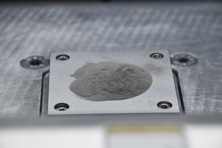
The nanoparticle-functionalized powder is fed into a 3D printer, which layers the powder and laser-fuses each layer to construct a 3D object.
Metals have not yet reached their 3D-printing potential. Of the more than 5,500 alloys used in manufacturing today, many cannot undergo the laser-based melting and quick cooling times without cracking or other defects.

Now researchers at HRL Laboratories, in Malibu, California have figured out that adding nanoparticles to the mix could prevent 3D-printed metals from cracking. Bringing metals into the additive manufacturing arena could lead to innovations in industries from aerospace to biomedicine by improving on conventional casting methods. Using 3D printing also allows products to be customized in a short period of time, making them available for sale more quickly.

“What we’re hoping to do is remove one of the constraints,” John Martin, a researcher at HRL Labs, told Seeker. “If you don’t have to worry about whether the material is going to be strong enough once you produce a part, you can focus on the next step, which is opening up the ideas of young engineers, who are thinking about brand new [design] geometries.”

In lab experiments the researchers 3D-printed two different kinds of aluminum alloys that not only didn’t crack, but were as strong as steel and one-third the weight. Martin and his colleagues published their research findings in the journal Nature.

Martin, who has studied metallurgy, knew the problem was at the atomic level. Metals are crystals and their atoms line up in repeating patterns that make up grains. There are sometimes defects at the boundaries where grains meet up and those defects can cause cracks.

There was also the issue of powdered metal. The process of 3D-printing metal requires that the metal begins as a powder, which is laid down in very thin layers, each one heated with a laser in order to melt it. The molten metal must cool quickly before the next layer of powder is added and laser-heated. The intense heating and quick cooling causes metal grains to solidify in odd shapes, which can lead to cracks.

Because the grains of powder are tiny and measured in microns, Martin and his team thought they could introduce nanosized particles to beef up the metal’s strength. They used a computer program to sort through and analyze more than 4,500 different alloy and nanoparticle combinations to see which ones had atomic structures that would fit together best. The idea was that the grains of metal would glom onto the tiny nanoparticles, sort of like a water vapor droplet that nucleates around a particle of dust to create a drop of rain.

They found that a nanoparticle made of hydrogen-stabilized zirconium would work best with two different kinds of aluminum alloys. During the very first lab experiment, they laid down a thin powdery layer of metal microparticles coated with a very fine layer of nanoparticles before running the laser over it. Layer after layer, the printer created the object until it was finished.

When it was completed, the scientists cut the object in half, then polished it. There were no cracks.

“I was surprised that it worked the first time,” said Martin. “We followed physics, so I wasn’t too shocked that it worked, but it’s easy to write something down on a piece of paper. It’s much harder to follow through.”

Read more at Seeker

Jul 19, 2017

Nanoparticles could spur better LEDs, invisibility cloaks

LED light bulb.
In an advance that could boost the efficiency of LED lighting by 50 percent and even pave the way for invisibility cloaking devices, a team of University of Michigan researchers has developed a new technique that peppers metallic nanoparticles into semiconductors.

It's the first technique that can inexpensively grow metal nanoparticles both on and below the surface of semiconductors. The process adds virtually no cost during manufacturing and its improved efficiency could allow manufacturers to use fewer semiconductors in finished products, making them less expensive.

The metal nanoparticles can increase the efficiency of LEDs in several ways. They can act as tiny antennas that alter and redirect the electricity running through the semiconductor, turning more of it into light. They can also help reflect light out of the device, preventing it from being trapped inside and wasted.

The process can be used with the gallium nitride that's used in LED lighting and can also boost efficiency in other semiconductor products, including solar cells. It's detailed in a study published in the Journal of Applied Physics.

"This is a seamless addition to the manufacturing process, and that's what makes it so exciting," said Rachel Goldman, U-M professor of materials science and engineering, and physics. "The ability to make 3-D structures with these nanoparticles throughout is going to open a lot of possibilities."

The key innovation


The idea of adding nanoparticles to increase LED efficiency is not new. But previous efforts to incorporate them have been impractical for large-scale manufacturing. They focused on pricey metals like silver, gold and platinum. In addition, the size and spacing of the particles must be very precise; this required additional and expensive manufacturing steps. Furthermore, there was no cost-effective way to incorporate particles below the surface.

Goldman's team discovered a simpler way that integrates easily with the molecular beam epitaxy process used to make semiconductors. Molecular beam epitaxy sprays multiple layers of metallic elements onto a wafer. This creates exactly the right conductive properties for a given purpose.

The U-M researchers applied an ion beam between these layers -- a step that pushes metal out of the semiconductor wafer and onto the surface. The metal forms nanoscale particles that serve the same purpose as the pricey gold and platinum flecks in earlier research. Their size and placement can be precisely controlled by varying the angle and intensity of the ion beam. And applying the ion beam over and over between each layer creates a semiconductor with the nanoparticles interspersed throughout.

"If you carefully tailor the size and spacing of nanoparticles and how deeply they're embedded, you can find a sweet spot that enhances light emissions," said Myungkoo Kang, a former graduate student in Goldman's lab and first author on the study. "This process gives us a much simpler and less expensive way to do that."

Researchers have known for years that metallic particles can collect on the surface of semiconductors during manufacturing. But they were always considered a nuisance, something that happened when the mix of elements was incorrect or the timing was off.

"From the very early days of semiconductor manufacturing, the goal was always to spray a smooth layer of elements onto the surface. If the elements formed particles instead, it was considered a mistake," Goldman said. "But we realized that those 'mistakes' are very similar to the particles that manufacturers have been trying so hard to incorporate into LEDs. So we figured out a way to make lemonade out of lemons."

Toward invisibility cloaks


Because the technique allows precise control over the nanoparticle distribution, the researchers say it may one day be useful for cloaks that render objects partially invisible by inducing a phenomenon known as "reverse refraction."

Reverse refraction bends light waves backwards in a way that doesn't occur in nature, potentially directing them around an object or away from the eye. The researchers believe that by carefully sizing and spacing an array of nanoparticles, they may be able to induce and control reverse refraction in specific wavelengths of light.

"For invisibility cloaking, we need to both transmit and manipulate light in very precise ways, and that's very difficult today," Goldman said. "We believe that this process could give us the level of control we need to make it work."

Read more at Science Daily

Mar 4, 2017

Most complex nanoparticle crystal ever made by design

The most complex crystal designed and built from nanoparticles. Left: An electron microscope image of a slice of the structure (Northwestern University). Right: A matching slice from a simulation of the structure (University of Michigan).
The most complex crystal designed and built from nanoparticles has been reported by researchers at Northwestern University and the University of Michigan. The work demonstrates that some of nature's most complicated structures can be deliberately assembled if researchers can control the shapes of the particles and the way they connect using DNA.

"This is a tour de force demonstration of what is possible when one harnesses the chemistry of DNA and combines it with nanoparticles whose shapes encourage a particular crystal structure," said Chad A. Mirkin, the George B. Rathmann Professor of Chemistry in the Weinberg College of Arts and Sciences at Northwestern.

Nanotechnology promises to bring materials together in new ways, forging new capabilities by design. One potential application for crystals built of nanoparticles, such as these newly reported ones, is the control of light -- nanoparticles interact well with light waves because they are similar in size. This could lead to materials that can change colors or patterns on command or block certain wavelengths of light, while transmitting or amplifying others. New types of lenses, lasers and even Star Trek-like cloaking materials are possible.

"This work shows that nanoparticle crystals of extraordinary complexity are possible with DNA technology, once one begins to exploit particle shape," said Sharon C. Glotzer, the John W. Cahn Distinguished University Professor of Engineering and the Stuart W. Churchill Collegiate Professor of Chemical Engineering at U-M. "And, it's a great example of what can be achieved by experimentalists and simulators teaming up."

The study, titled "Clathrate Colloidal Crystals," will be published March 3 in the journal Science. Mirkin and Glotzer are co-corresponding authors of the paper.

In chemistry, clathrates are known for their chambers that can house small molecules. They have been used for capturing pollutants from the environment, for example. The nanoparticle clusters also leave room for cargo, which the authors suggest could be useful for storing, delivering and sensing materials for environmental, medical diagnostic and therapeutic applications.

While natural materials exhibit a dizzying array of crystal structures, most nanotechnology labs struggle to get past simple designs. The structures produced by Haixin Lin, now a postdoctoral fellow in Mirkin's lab, are far more interesting. The new structures are composed of clusters of up to 42 particles, forming larger polyhedral, such as the great dodecahedron. These clusters connect into cage-like crystal structures called clathrates.

Still, the story isn't the crystal itself: it's how the crystal came to be. Mirkin's group has pioneered many structures through the use of DNA strands as a sort of smart glue, linking nanoparticles together in a particular way. The particle is both a building block and a template that directs bonding interactions. Meanwhile, Glotzer's group has championed the role of nanoparticle shape in guiding the assembly of crystal structures through computer simulation.

"Chad's group got the idea of exploring new phases by looking at predictions we had made," Glotzer said. "One day, I got a phone call from him. 'We just got these incredible structures!' he said. And he texted me micrograph after micrograph -- they just kept popping up. He said, we need to figure out a way to definitively assign their structures."

The electron microscope images, or micrographs, showed complex crystalline structures that formed in large part thanks to the shape of the gold nanoparticles. The triangular bipyramidal shape, like two flattened tetrahedrons stuck together at their bases, was similar to a shape Glotzer's group predicted would form a quasicrystal. Quasicrystals are prized in the field of nanoassembly because they are as complex as crystals get.

Lin's shape had just the right angles to make clathrate structures, which often turn up in molecular systems that form quasicrystals. But to do so, they needed strands of DNA attached to their sides at just the right length.

Lin systematically made the gold bipyramids of consistent size and shape, with edge lengths of 250 nanometers -- half the wavelength of blue light. He then modified them with different length sequences of DNA. When the DNA strands were too short, the nanoparticles made disordered, ill-defined structures.

When longer strands produced exotic patterns in the electron microscope images, Lin brought the results to Mirkin, who was both thrilled and intrigued.

"These are stunning -- no one has made such structures before," said Mirkin, director of Northwestern's International Institute for Nanotechnology.

It was clear they had made phases never observed before, but getting the structure accurately identified was essential. After Mirkin alerted Glotzer at U-M, Sangmin Lee and Michael Engel 3-D printed Lin's bipyramids and glued them together to explore how they might make the structures in the electron micrographs. Lee is a doctoral student in chemical engineering, and Engel was then an assistant research scientist, both in Glotzer's group.

Once they saw how the shapes fit together, they hypothesized the clathrate structures. To confirm their suspicions, they built a computer model of the hypothesized clathrates from bipyramids and compared it to the Northwestern micrographs. It was a perfect match.

As a definitive test, Lee and Matthew Spellings, also a doctoral student in chemical engineering at U-M, developed a molecular model of the DNA-linked nanoparticles. Lee carried out simulations to confirm that the particles would indeed form clathrate structures.

"To really know for sure, we had to run simulations that mimicked the conditions Haixin used in the lab to see if a disordered fluid of DNA-linked bipyramids would assemble into the Northwestern crystals," Glotzer said. "Once we saw the computer crystals, I knew we had nailed it."

Mirkin is director of the research group that invented the chemistry for conjugating DNA and nanoparticles and a pioneer of the concept of programmable colloidal crystallization with nucleic acids. In 1996, he introduced the concept of using nanoparticles as atoms and synthetic DNA -- the blueprint of life -- as a chemically programmable bond to make designer materials based upon the ability of the particles to recognize one another through sequences immobilized on their surfaces. Mirkin has been experimentally studying the role of particle shape in colloidal crystal formation for the past two decades. He is also a professor of medicine, chemical and biological engineering, biomedical engineering, and materials science and engineering.

Read more at Science Daily

Mar 12, 2016

'Sticky waves: ' molecular interactions at the nanoscale

Like the gravitational forces that are responsible for the attraction between the Earth and the moon, as well as the dynamics of the entire solar system, there exist attractive forces between objects at the nanoscale.

These are the so-called van der Waals forces, which are ubiquitous in nature and thought to play a crucial role in determining the structure, stability and function of a wide variety of systems throughout the fields of biology, chemistry, physics and materials science.

"To put it simply, every molecular system and every material in nature experiences these forces," said Robert A. DiStasio Jr., assistant professor of chemistry and chemical biology in the College of Arts and Sciences. "In fact, we are finding that their influence is quite extensive, and includes protein-drug interactions, the stability of the DNA double helix, and even the peculiar adhesion properties of the gecko's foot."

When compared with the covalent bond (which involves the sharing of electron pairs between atoms), van der Waals forces are relatively weak and arise from instantaneous electrostatic interactions between the fluctuating electron clouds that surround microscopic objects. However, these forces are still quantum mechanical in origin and have posed a substantial challenge for both theory and experiment to date.

In a paper in the March 11 issue of Science, DiStasio and collaborator Alexandre Tkatchenko of the University of Luxembourg and the Fritz Haber Institute have put forth a new proposition for describing van der Waals forces among objects at the nanoscale.

Generally speaking, there are two schools of thought regarding these forces. The prevailing description of van der Waals interactions among most chemists and biologists is the picture of two induced electric dipoles, similar to the N and S poles of a magnet, representing the uneven distributions of positive and negative charges. The picture espoused by many physicists, however, centers around the fact that wavelike vacuum fluctuations are responsible for the van der Waals interactions among larger macroscopic objects.

In their work, DiStasio and Tkatchenko demonstrate that these fundamental forces between nanostructures must also be described by the electrostatic interactions between wavelike (or delocalized) charge density fluctuations instead of the aforementioned particle-like (or local) induced dipoles. They believe their work could help to bridge the gap between these two belief systems, and help scientists understand and control the interactions between objects at the nanoscale.

"Our work is demonstrating that there is a much wider variety of systems, such as nanostructured systems, where you have to think about the van der Waals force in terms of interactions between waves instead of interactions between particles," Tkatchenko said.

Paul McEuen, the John A. Newman Professor of Physical Science and director of the Kavli Institute at Cornell for Nanoscale Science, sees the duo's research as an important first step in a long, complicated journey to what McEuen half-jokingly characterized as "solving biology."

"It sounds like a rather boring problem, but it's actually a deeply important problem, the way biomolecules assemble and so on," said McEuen. "It's a hugely important problem, especially for someone like me, who's a nano-guy, but it's going to take time to solve."

Read more at Science Daily

Feb 27, 2016

The key to mass-producing nanomaterials

Nanoparticles -- tiny particles 100,000 times smaller than the width of a strand of hair -- can be found in everything from drug delivery formulations to pollution controls on cars to HD TV sets. With special properties derived from their tiny size and subsequently increased surface area, they're critical to industry and scientific research.

They're also expensive and tricky to make.

Now, researchers at USC have created a new way to manufacture nanoparticles that will transform the process from a painstaking, batch-by-batch drudgery into a large-scale, automated assembly line.

The method, developed by a team led by Noah Malmstadt of the USC Viterbi School of Engineering and Richard Brutchey of the USC Dornsife College of Letters, Arts and Sciences, was published in Nature Communications on Feb. 23.

Consider, for example, gold nanoparticles. They have been shown to be able to easily penetrate cell membranes without causing any damage -- an unusual feat, given that most penetrations of cell membranes by foreign objects can damage or kill the cell. Their ability to slip through the cell's membrane makes gold nanoparticles ideal delivery devices for medications to healthy cells, or fatal doses of radiation to cancer cells.

However, a single milligram of gold nanoparticles currently costs about $80 (depending on the size of the nanoparticles). That places the price of gold nanoparticles at $80,000 per gram -- while a gram of pure, raw gold goes for about $50.

"It's not the gold that's making it expensive," Malmstadt said. "We can make them, but it's not like we can cheaply make a 50 gallon drum full of them."

Right now, the process of manufacturing a nanoparticle typically involves a technician in a chemistry lab mixing up a batch of chemicals by hand in traditional lab flasks and beakers.

Brutchey and Malmstadt's new technique instead relies on microfluidics -- technology that manipulates tiny droplets of fluid in narrow channels.

"In order to go large scale, we have to go small," Brutchey said. Really small.

The team 3D printed tubes about 250 micrometers in diameter -- which they believe to be the smallest, fully enclosed 3D printed tubes anywhere. For reference, your average-sized speck of dust is 50 micrometers wide.

They then built a parallel network of four of these tubes, side-by-side, and ran a combination of two non-mixing fluids (like oil and water) through them. As the two fluids fought to get out through the openings, they squeezed off tiny droplets. Each of these droplets acted as a micro-scale chemical reactor in which materials were mixed and nanoparticles were generated. Each microfluidic tube can create millions of identical droplets that perform the same reaction.

Read more at Science Daily