Showing posts with label Biotechnology. Show all posts

Jan 28, 2021

Making wheat and peanuts less allergenic

 The United States Department of Agriculture identifies a group of "big eight" foods that causes 90% of food allergies. Among these foods are wheat and peanuts.

Sachin Rustgi, a member of the Crop Science Society of America, studies how we can use breeding to develop less allergenic varieties of these foods. Rustgi recently presented his research at the virtual 2020 ASA-CSSA-SSSA Annual Meeting.

Allergic reactions caused by wheat and peanuts can be prevented by avoiding these foods, of course. "While that sounds simple, it is difficult in practice," says Rustgi.

Avoiding wheat and peanuts means losing out on healthy food options. These two foods are nutritional powerhouses.

Wheat is a great source of energy, fiber, and vitamins. Peanuts provide proteins, good fats, vitamins and minerals.

"People with food allergies can try hard to avoid the foods, but accidental exposure to an allergen is also possible," says Rustgi. Allergen exposure can lead to hospitalization, especially for people with peanut allergies.

"For others, avoiding wheat and peanuts is not easy due to geographical, cultural, or economic reasons," explains Rustgi.

Rustgi and his colleagues are using plant breeding and genetic engineering to develop less allergenic varieties of wheat and peanuts. Their goal is to increase food options for people with allergies.

For wheat, researchers focus on a group of proteins, called gluten.

The gluten in bread flour makes dough elastic. Gluten also contributes to the chewy texture of bread.

But gluten can cause an immune reaction for individuals with Celiac disease. In addition, others experience non-celiac gluten sensitivity, leading to a variety of adverse symptoms.

Researchers have been trying to breed varieties of wheat with lower gluten content. The challenge, in part, lies in the complicated nature of gluten genetics. The information needed to make gluten is embedded in the DNA in wheat cells.

But gluten isn't a single protein -- it's a group of many different proteins. The instructions cells needed to make the individual gluten proteins are contained within different genes.

In wheat, these gluten genes are distributed all over a cell's DNA. Since so many portions of the DNA play a role in creating gluten, it is difficult for plant breeders to breed wheat varieties with lower gluten levels.

"When we started this research, a major question was whether it would be possible to work on a characteristic controlled by so many genes," says Rustgi.

For peanuts, the situation is similar. Peanuts contain 16 different proteins recognized as allergens.

"Not all peanut proteins are equally allergenic," says Rustgi. Four proteins trigger an allergic reaction in more than half of peanut sensitive individuals.

Like the gluten genes in wheat, the peanut allergen genes are spread throughout the peanut DNA.

"Affecting this many targets is not an easy task, even with current technology," says Rustgi.

Rustgi and the research team are testing many varieties of wheat and peanuts to find ones that are naturally less allergenic than others.

These low-allergenic varieties can be bred with crop varieties that have desirable traits, such as high yields or pest resistance. The goal is to develop low-allergenic wheat that can be grown commercially.

In addition to traditional breeding efforts, Rustgi is also using genetic engineering to reduce allergenic proteins in wheat and peanuts.

For example, a technology called CRISPR allows scientists to make very precise changes to a cell's DNA.

Rustgi is using CRISPR to target gluten genes in wheat. Recent improvements in CRISPR technology allow researchers to target many genes at once.

Genes targeted by CRISPR are changed or mutated. This means that cells can no longer 'read' these genes to make the specific proteins.

"Disrupting the gluten genes in wheat could yield wheat with significantly lower levels of gluten. A similar approach would work in peanuts," says Rustgi.

Other approaches include understanding how gluten production is regulated in wheat cells. As it turns out, one protein serves as a 'master regulator' for many gluten genes.

That's important because disrupting this master regulator could lead to reduced amounts of gluten in wheat. Targeting a single gene is much easier than trying to disrupt the several gluten genes.

"Wheat and peanuts are the major sources of proteins to many, especially those living in resource-deprived conditions," says Rustgi. "Finding affordable ways to make wheat and peanuts available for all is very important."

Developing wheat and peanuts with reduced allergen levels is a key step toward this goal.

Read more at Science Daily

Jan 24, 2018

Chinese Lab Clones the World's First Primates Using Dolly-the-Sheep Technique

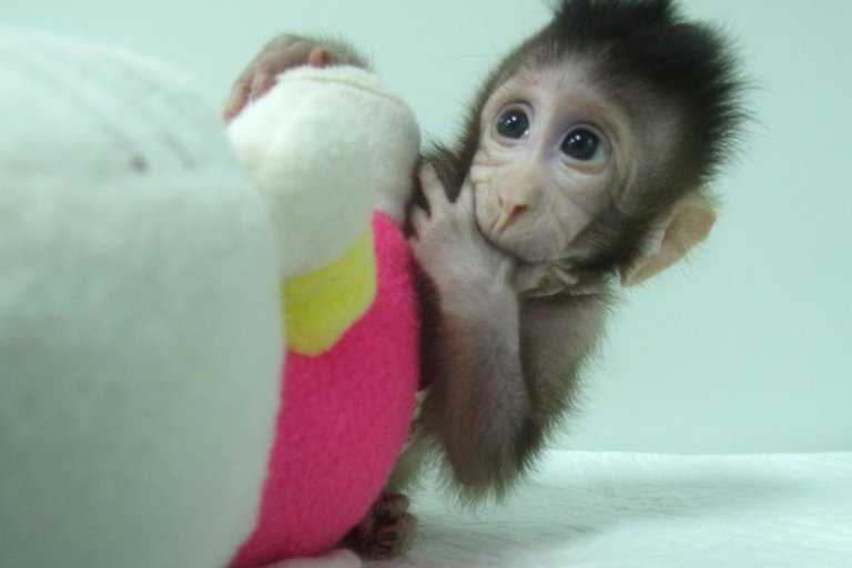
This is a photograph of Zhong Zhong, one of the first two monkeys created by somatic cell nuclear transfer.
Geneticists in China announced that they've successfully cloned the first primates using somatic cell nuclear transfer, the same method that produced Dolly, the first cloned sheep more than 20 years ago.

According to a new paper in the journal Cell, the procedure will it make possible for labs to decant large populations of genetically uniform monkeys, which could open up entirely new areas for medical research.

The long-tail macaques, named Zhong Zhong and Hua Hua, were born six and eight weeks ago, respectively. While the monkeys are not the first primates to be cloned — others have been created using a simpler process called embryo splitting — they are the first to ever survive the somatic cell nuclear transfer (SCNT) process. The advantage to this technique, researchers say, is that greater numbers of genetically identical clones can be created in a single batch. This allows geneticists to make isolated changes in the genetic code, then study outcomes among a large population.

“There are a lot of questions about primate biology that can be studied by having this additional model,” said senior author Qiang Sun, director of the Nonhuman Primate Research Facility at the Chinese Academy of Sciences Institute of Neuroscience, in a statement issued with the publication of the new research. "You can produce cloned monkeys with the same genetic background except the gene you manipulated. This will generate real models not just for genetically based brain diseases, but also cancer, immune, or metabolic disorders and allow us to test the efficacy of the drugs for these conditions before clinical use."

This is a photograph of Hua Hua, one of the first monkey clones made by somatic cell nuclear transfer.
When using the SCNT process, scientists remove the nucleus of an egg cell and replace it with another nucleus, which can in turn be taken from various tissue types of either fetal or adult donor specimens. In the case of the Chinese macaques, researchers took fibroblasts — a cell type found in connective tissue — from a single, fetal macaque. As such, the two new monkeys are exact clones of each other, as well as the original specimen.

By comparison, the embryo splitting technique — the one that had been previously used with monkeys — is essentially an artificial approximation of how natural twins develop in mammals. Embryo splitting can only generate up to four offspring at a time, while SCNT clones are theoretically limitless.

In either case, cloned egg cells are placed within surrogate female monkey moms, where the embryos develop and are born naturally.

The research team had previously tried to use adult donor cells with the SCNT method, but the cloned macaques died a few hours after birth. Earlier research established that, for reasons that are still unclear, monkey cell nuclei are more resistant to the SCNT process than other mammals, such as mice and cows.

"We tried several different methods, but only one worked," said Sun. "There was much failure before we found a way to successfully clone a monkey."

The researchers plan to continue improving the technique and generating more macaque clones, using international guidelines for animal research set by the US National Institutes of Health.

“We are very aware that future research using non-human primates anywhere in the world depends on scientists following very strict ethical standards,” said Muming Poo, a co-author of the study.

Read more at Seeker

May 26, 2017

Living Cell Circuits Could Regrow Organs and Produce Biofuels

An artist's impression of connected CRISR-dCas9 NOR gates.
It's no secret that computer circuits can process enormous amounts of information — entire industries are built around this core function of technology.

But biologists will tell you that living cells process information too, and their process isn't a simple binary exchange of ones and zeros. Instead, cells deal with multiple complex structures of sugars, proteins, lipids, and DNA. As such, the act of “programming” cells, through genetic engineering, is enormously complex.

But that's okay — scientists like a good challenge.

In research from the University of Washington, published in the journal Nature Communications, synthetic biologists announced a new method for turning organic cells into living computers. By installing the organic equivalent of the digital logic gates used in electronics, scientists can code instructions into the cell so that particular inputs result in desired outputs. Instead of silicon and solder, biologists are using DNA and yeast cells to developing this new kind of organic processor.

In a series of experiments, the UW team built the largest organic “circuit board” constructed to date, including seven logic gates assembled in series or parallel.

Each gate consists of a gene with three programmable chunks of DNA. Two act as inputs, with the third as the output. Using CRISPR technology, the researchers programmed specific proteins to act as molecular gatekeepers, determining whether a particular gate will be active or not.

If a gate is active, it sends a signal that deactivates another gate within the organic circuit, which means scientists can basically “wire” together the gates to create logical programs in the cell.

The new research is a significant step forward for synthetic biology, said senior author and UW electrical engineering professor Eric Klavins.

“Digital logic has been done at a small scale, a few gates, many times over the last decade or so,” Klavins said in an email. “Our paper is the first to produce large circuits built in a eukaryote (yeast), in which transcriptional machinery is considerable more complex.”

Eurkaryotic cells, like human cells, contain a nucleus and other structures that enable complex behaviors. These are the kinds of cells we'll need to hack for doing those really useful things — like regrow a liver.

“Cells could be reprogrammed to undergo new developmental pathways, to regrow organs, or to develop entirely new ones,” Klavins said. “In such developing tissues, cells have to make complex digital decisions about what genes to express and when, and our technology could be used to control that process.”

The organic circuit board could also be used to produce viable biofuels, which requires importing genes from several different organisms into a hybrid industrial strain, Klavins said.

“Those genes express protein enzymes that each do a particular conversion of one molecule to another, along a metabolic pathway that usually starts with sugar and ends with the fuel,” he said.

Read more at Discovery News

Nov 9, 2016

Brain Implant Could Help Paraplegics Walk Again

A groundbreaking new technology unveiled this week could enable paraplegics to walk again by sending signals wirelessly from the brain to the lower part of the spine.

The neuroprosthetic interface, developed by an international team of scientists and technicians, acts as bridge between the brain's motor cortex and the spine. Brain signals are picked up by an implant in the skull and then translated by a small computer, which relays the information — wirelessly and immediately — to electrodes implanted in the spine.

The spinal electrodes are placed in the lumbar region, below the injury or lesion that otherwise prevents signals from passing through the nerves. The electrodes, in turn, stimulate the neural pathways that activate leg muscles when walking or running. The interface essentially creates a wireless connection between the brain and the spine, potentially allowing those with spinal injuries to walk again.

So far, the system has only been tested on monkeys, but initial results indicate that the system could work with humans, according to the research team.

David Borton, assistant professor of engineering with Brown University and co-author of the new study, said that spinal stimulation studies with humans are being evaluated, but there are no clinical trials in the works just yet.

"The current paper shows encouraging evidence that such brain recording and spinal stimulation could be used in a human rehabilitation context, but no studies are currently planned," Brown said in an email to Seeker.

According to the study, officially published today in the journal Nature, two rhesus monkeys were outfitted with the neuroprosthetic earlier this year. Both monkeys had one leg paralyzed by a spinal cord lesion. On June 23, 2015, the first monkey regained control of its paralyzed leg, less than one week after the initial injury. The second monkey recovered within two weeks. Both animals were able to walk again, without training, both on the ground and on treadmills.

The neuroprosthetic interface was conceived at EPFL in Switzerland, and developed together with an international network of collaborators including Irish medical device company Medtronic, Brown University and the German research institution Fraunhofer ICT-IMM. Testing was performed in collaboration with the University of Bordeaux, Motac Neuroscience and the Lausanne University Hospital (CHUV).

As you might imagine, the technical details are complex (you can peruse the research paper itself at Nature) but the system basically consists of five components: the brain implant, a brain-recording device, a computer, an implantable pulse generator and a spinal implant.

The brain implant technology has previously been deployed to control movement in prosthetic hands and, in one case, a patient's fully paralyzed hand. But this is the first time it's been used for spinal injuries.

The computer element of the system extracts specific intentional information from the brain implant, then translates the data using special algorithms. The data is beamed wirelessly to the pulse generator, which delivers the instructed patterns of simulation to the spinal implant. The spinal implant activates groups of muscles in the paralyzed leg, inducing flexing and extension movements.

Read more at Discovery News

Jul 31, 2016

Novel 'repair system' discovered in algae may yield new tools for biotechnology

This is a TEM image of the algae C. reinhardtii.
A new way of fixing inactive proteins has been discovered in an algae, which uses chloroplast extracts and light to release an interrupting sequence from a protein.

Research specialist Stephen Campbell and Professor David Stern at the Boyce Thompson Institute report the discovery in the July 29 issue of the Journal of Biological Chemistry. This repair system may have applications in agriculture and biotechnology because it could potentially be harnessed to enable proteins to become active only in the light.

Many proteins contain extra sequences, called insertions, that can disrupt their function. The current paper demonstrates that the algae Chlamydomonas reinhardtii has the necessary toolkit to repair proteins by removing these insertions.

Campbell discovered this new repair system while purifying a protein from the chloroplasts of C. reinhardtii that can cut RNA. Upon sequencing the protein, he identified it as RB47, a protein that was not known to have any RNA-cleaving ability. Campbell noticed that the middle of the protein was missing. When he compared the protein sequence to its corresponding gene sequence, the protein was much shorter than expected.

Upon further study, Campbell found that he could detect a long version of the protein that contained an insertion and a short version that didn't. The cells make both versions when grown in the light or the dark, but only the short version can cleave RNA. The long version of the protein could be converted into the short one by mixing it in a test tube with chloroplasts from cells grown in the light and by illuminating the reaction. This process removed the interrupting insertion and restored the RNA-cutting activity of the protein. It is likely that the chloroplast maintains the machinery necessary to remove the sequence so that it can restore functionality to the protein.

This new type of repair system provides intriguing possibilities for biotech applications.

Because the insertion can be placed so that it interrupts a protein's function, the insertion and repair system may be useful for producing certain pharmaceuticals or protein products -- such as cancer drugs -- in culture, which would otherwise kill the cell. After purification, the inactive products could be treated with chloroplast factors and light to remove the insertion and activate the proteins.

In future work, the researchers plan to investigate exactly how the insertion becomes spliced out of the protein and which plant factors facilitate its removal. They also aim to understand the purpose of the insertion, and whether the algae can control the splicing to respond to changes in the environment.

Read more at Science Daily

Sep 13, 2014

Worldwide study demonstrates accuracy of genetic analyses

Physicians envision a future in which genomic data from patients is heavily used to manage care, but experts have questioned the accuracy and reliability of these analyses. Now, a study by 150 researchers in 12 countries finds real strength and agreement across RNA genomic sequencing techniques and laboratories -- as well as ways to improve what little variability exists to set a new high standard.

The results of the study were published in Nature Biotechnology in three separate research articles.

These results should provide assurance to patients, clinicians and the research community that genomic sequencing is accurate, says E. Aubrey Thompson, Ph.D., a professor of cancer biology at Mayo Clinic in Florida, one of three institutions that led the study. Dr. Thompson is a study co-author and member of the project leadership.

"It seems very likely that decisions about patient care are going to be influenced by genomic data, derived from sequencing both RNA and DNA from patient samples, and we now know the extent to which these sequence-based analyses can be relied upon within a given laboratory or from laboratory to laboratory," he says.

"That means that results of a patient's sample, from which clinical management decisions will likely be made, will be accurate worldwide," says Dr. Thompson.

RNA sequencing is being used with increasing frequency to characterize a growing array of conditions -- everything from prenatal birth defects to disorders of the elderly.

The other institutions involved in the study are the Beijing Genomic Institute and Weill Cornell Medical School. All three institutions have extensive experience in sequencing RNA and have helped develop novel analytical tools for interpreting the data.

The U.S. Food and Drug Administration (FDA) funded the research, given its need to understand the accuracy of such data submitted in applications for approval of new drugs, clinical applications and genomic diagnostic procedures, Dr. Thompson says.

The purpose of this project, known as Sequence Quality Control (SEQC), was to rigorously define both the scope and the sources of variation in RNA sequencing data.

Laboratory groups at the three leading institutions sequenced the same two RNA samples multiple times.

More than 1 billion nucleotides of sequencing data were generated by each site. The data were then analyzed under the direction of the FDA with the assistance of a large group of academic and industrial statisticians. The researchers also examined the current technologies and major biochemical methods of 30 RNA-sequencing labs and hundreds of researchers. The researchers also found that RNA can be accurately extracted and analyzed from severely degraded genetic samples, such as from tissue samples that have been stored for many years.

Read more at Science Daily

May 31, 2014

3-D bioprinting builds a better blood vessel

The tangled highway of blood vessels that twists and turns inside our bodies, delivering essential nutrients and disposing of hazardous waste to keep our organs working properly has been a conundrum for scientists trying to make artificial vessels from scratch. Now a team from Brigham and Women's Hospital (BWH) has made headway in fabricating blood vessels using a three-dimensional (3D) bioprinting technique.

The study is published online this month in Lab on a Chip.

"Engineers have made incredible strides in making complex artificial tissues such as those of the heart, liver and lungs," said senior study author, Ali Khademhosseini, PhD, biomedical engineer, and director of the BWH Biomaterials Innovation Research Center. "However, creating artificial blood vessels remains a critical challenge in tissue engineering. We've attempted to address this challenge by offering a unique strategy for vascularization of hydrogel constructs that combine advances in 3D bioprinting technology and biomaterials."

The researchers first used a 3D bioprinter to make an agarose (naturally derived sugar-based molecule) fiber template to serve as the mold for the blood vessels. They then covered the mold with a gelatin-like substance called hydrogel, forming a cast over the mold which was then reinforced via photocrosslinks.

"Our approach involves the printing of agarose fibers that become the blood vessel channels. But what is unique about our approach is that the fiber templates we printed are strong enough that we can physically remove them to make the channels," said Khademhosseini. "This prevents having to dissolve these template layers, which may not be so good for the cells that are entrapped in the surrounding gel."

Khademhosseini and his team were able to construct microchannel networks exhibiting various architectural features. They were also able to successfully embed these functional and perfusable microchannels inside a wide range of commonly used hydrogels, such as methacrylated gelatin or poly(ethylene glycol)-based hydrogels at different concentrations.

Read more at Science Daily

May 15, 2014

Why Octopuses Don't Tie Themselves in Knots

Octopuses look like a tangle of squishy arms, and yet they never tie their limbs in a knot, finds a new study that determined octopus skin repels itself.

The discovery, published in the latest issue of Current Biology, is helping researchers to design soft robots, such as for surgical use, that can reshape their bodies without becoming a jumbled mess.

It also solves a mystery about octopuses, whose brains appear to be are unaware of what their two legs and six arms are doing. Instead, a chemical produced by octopus skin temporarily prevents octopus suckers from sucking.

"We were surprised that nobody before us had noticed this very robust and easy-to-detect phenomena," co-author Guy Levy of the Hebrew University of Jerusalem, said in a press release. "We were entirely surprised by the brilliant and simple solution of the octopus to this potentially very complicated problem."

We humans don't have such problems because our rigid skeletons limit the number of possibilities as to where our arms and legs could be.

"Our motor control system is based on a rather fixed representation of the motor and sensory systems in the brain in a formant of maps that have body part coordinates," explained co-author Binyamin Hochner.

He continued, "It is hard to envisage similar mechanisms to function in the octopus brain because its very long and flexible arms have an infinite number of degrees of freedom. Therefore, using such maps would have been tremendously difficult for the octopus, and maybe even impossible."

As a demonstration of that freedom, check out this video that shows just how amazingly flexible these marine animals can be:

For the study, Hochner and his colleagues observed the behavior of amputated octopus arms, which remain very active for an hour after separation. (Octopuses can grow their arms back, but doing so is energetically costly.)

Those observations showed that the arms never grabbed octopus skin, though they would grab a skinned octopus arm. The octopus arms didn’t grab Petri dishes covered with octopus skin either, and they attached to dishes covered with octopus skin extract with much less force than they otherwise would.

"The results so far show, and for the first time, that the skin of the octopus prevents octopus arms from attaching to each other or to themselves in a reflexive manner," the researchers wrote. "The drastic reduction in the response to the skin crude extract suggests that a specific chemical signal in the skin mediates the inhibition of sucker grabbing."

Read more at Discovery News

Nov 30, 2013

Researchers Find Missing Component in Effort to Create Primitive, Synthetic Cells

A team of Massachusetts General Hospital (MGH) investigators working to create "protocells" -- primitive synthetic cells consisting of a nucleic acid strand encased within a membrane-bound compartment -- have accomplished an important step towards their goal. In the November 28 issue of Science, the investigators describe a solution to what could have been a critical problem -- the potential incompatibility between a chemical requirement of RNA copying and the stability of the protocell membrane.

"For the first time, we've been able to do nonenzymatic RNA copying inside fatty acid vesicles," says Jack Szostak, PhD, of the MGH Department of Molecular Biology and the Center for Computational and Integrative Biology. "We've found a solution to a longstanding problem in the origin of cellular life: RNA copying chemistry requires the presence of the magnesium ion Mg2+, but high Mg2+ levels can break down the simple, fatty acid membranes that probably surrounded the first living cells."

Szostak's team has been working for more than a decade to understand how the first cells developed from a "primordial soup" of chemicals into living organisms capable of copying their genetic material and reproducing. Part of that work is developing a model protocell made from components probably present in the primitive Earth environment. They have made significant progress towards developing cell membranes from the kind of fatty acids that would have been abundant and naturally form themselves into bubble-like vesicles when concentrated in water. But the genetic component -- an RNA or DNA molecule capable of replication -- has been missing.

Since the primitive environment in which such cells could have developed would not have had the kind of complex enzymes that modern cells use in replicating nucleic acids, Szostak and lead author Katarzyna Adamala, PhD, then a graduate student in Szostak's lab, investigated whether simple chemical processes could drive nonenzymatic replication of RNA, which many scientists believe was the first nucleic acid to develop.

To address the incompatibility between the need for Mg2+ to drive assembly of the RNA molecule and the ion's ability to degrade fatty acid membranes, they tested several chelators -- small molecules that bind tightly to metal ions -- for their ability to protect fatty acid vesicles from the potentially destabilizing effects of Mg2+. Citrate and several other chelators were found to be effective in protecting the membranes of fatty acid vesicles from disruption.

To test whether the presence of the tested chelators would allow Mg2+-catalyzed RNA assembly, the investigators placed molecules consisting of short primer RNA strands bound to longer RNA templates into fatty acid vesicles. The unbound, single-strand portion of the template consisted of a sequence of cytosine (C) nucleotides. In the presence of Mg2+ and one of four chelating molecules, one of which was citrate, the researchers then added activated G, the nucleotide that base-pairs with C in nucleic acids.

The desired reaction -- diffusion of G nucleotides through the vesicle membrane to complete a double-stranded RNA molecule by binding to the C nucleotides of the template -- proceeded fastest in the presence of citrate. In fact two of the other tested chelators completely prevented extension of the RNA primer.

"While other molecules can protect membranes against the magnesium ion," Szostak explains, "they don't let RNA chemistry go on. We think that citrate is able both to protect membranes and to allow RNA copying to proceed by covering only one face to the magnesium ion, protecting the membrane while allowing RNA chemistry to work." He and Adamala also found that continually refreshing the activated guanine nucleotide solution by flushing out broken down molecules and adding fresh nucleotides improved the efficiency of RNA replication.

Read more at Science Daily

Nov 28, 2013

Scientists Stitch Up Photosynthetic Megacomplex

When sunlight strikes a photosynthesizing organism, energy flashes between proteins just beneath its surface until it is trapped as separated electric charges. Improbable as it may seem these tiny hits of energy eventually power the growth and movement of all plants and animals. They are literally the sparks of life.

The three clumps of protein -- a light-harvesting antenna called a phycobilisome and photosystems I and II -- look like random scrawls in illustrations but this is misleading. They are able to do their job only because they are positioned with exquisite precision.

If the distances between proteins were too great or the transfers too slow, the energy would be wasted and -- ultimately -- all entropy-defying assemblages like plants and animals would fall to dust.

But until now scientists weren't even sure the three complex cohered as a single sun-worshipping megacomplex. Previous attempts to isolated connected complexes failed because the weak links that held them together broke and the megacomplex fell apart.

In the Nov. 29 issue of Science scientists at Washington University in St. Louis report on a new technique that finally allows the megacomplex to be plucked out entire and examined as a functioning whole.

Like a seamstress basting together the pieces of a dress, the scientists chemically linked the proteins in the megacomplex. Stabilized by the stitches, or crosslinks, it was isolated in its complete, fully functional form and subjected to the full armamentarium of their state-of-the-art labs, including tandem mass spectrometers and ultra-fast lasers.

The work was done at PARC (Photosynthetic Antenna Research Center), an Energy Frontier Research Center funded by the Department of Energy that is focused on the scientific groundwork needed to maximize photosynthetic efficiency in living organisms and to design biohybrid or synthetic ones to drive chemical processes or generate photocurrent.

Robert Blankenship, PhD, PARC's director and the Lucille P. Markey Distinguished Professor of Arts & Sciences, said that one outcome of the work in the long term might be the ability to double or triple the efficiency of crop plants -- now stuck at a woeful 1 to 3 percent. "We will need such a boost to feed the 9 or 10 billion people predicted to be alive by 2050," he said.

Wizards of the lab

The scientists worked with the model organism often used to study photosynthesis in the lab, a cyanobacterium, sometimes called a blue-green alga.

Cyanobacteria are ancient organisms, known from fossils that are 3.5 billion years old, nearly as old as the oldest known rocks, and thought to be the first organisms to release oxygen into the noxious primitive atmosphere.

All photosynthesizing organisms have light-harvesting anntenas made up of many molecules that absorb light and transfer the excitation energy to reaction centers, where it is stored as charge separation.

In free-living cyanobacteria the antenna, called a phycobilisome, consists of splayed rods made up of disks of proteins containing intensely colored bilin pigments. The antenna sits directly above one reaction center, Photosystem II, and kitty corner to the other, Photosystem I.

PARC research scientist Haijun Liu, PhD, proposed stitching together the megacomplex and then engineered a strain of cyanobacteria that has a tag on the bottom of Photosystem II.

The mutant cells were treated with reagents that stitched together the complexes, then broken open, and the tag used to pull out Photosystem II and anything attached to it.

To figure out how the proteins were interconnected, the scientists repeatedly cut or shattered the proteins, analyzing them by mass spectrometry down to the level of the individual amino acid.

The amino acid sequences derived in this way were then compared to known sequences within the megacomplex, and the location of cross links between different complexes helped establish the overall structure of the megacomplex.

"It's a very complicated data analysis routine that literally generates tens of thousands of peptides that took a team of students and postdoctoral associates overseen by Hao Zhang and Michael Gross, months to analyze," Blankenship said. Hao Zhang, PhD, is a PARC research Scientist and Michael Gross, PhD, is professor of chemistry and Director of the Mass Spectrometry Resource in Arts & Sciences.

In the meantime research scientist Dariusz Niedzwiedzki, PhD, in the PARC Ultrafast Laser Facility was exciting the phycobilisome in intact megacomplexes and tracking the energy through the complex by the faint glow of fluorescencing molecules.

Typical energy transfers within the complex take place in a picosecond (a trillionth of a second), way too fast for humans to perceive. If one picosecond were a second, a second would be 31,700 years.

"PARC is one of the only places in the world that has available this sophisticated combination of experience and advanced techniques," said Blankenship, "and to solve this problem we were brought all of our expertise to bear.

"The work provides a new level of understanding of the organization of these photosynthetic membranes and that is something that a lot of people have tried to understand for a long time," he said.

Read more at Science Daily

Aug 28, 2013

Tiny Brain Parts Teased from Stem Cells

Ear, eye, liver, windpipe, bladder, and even a heart. The list of body parts grown from stem cells is getting longer and longer. Now add to it one of the most complex organs: the brain.

A team of European scientists has grown parts of a human brain in tissue culture from stem cells. Their work could help scientists understand the origins of schizophrenia or autism and lead to drugs to treat them, said Juergen Knoblich, deputy scientific director at the Institute of Molecular Biotechnology of the Austrian Academy of Science and one of the paper's co-authors.

The advance could also eliminate the need for conducting experiments on animals, whose brains are not a perfect model for humans.

To grow the brain structures, called organoids, the scientists used stem cells, which can develop into any other kind of cell in the body. They put the stem cells into a special solution designed to promote the growth of neural cells. Bits of gel interspersed throughout the solution gave the cells a three-dimensional structure to grown upon. In eight to ten days the stem cells turned into brain cells. After 20 days to a month, the cells matured into a size between three and four millimeters, representing specific brain regions, such as the cortex and the hindbrain.

Growing brain tissue this way marks a major advancement because the lab-grown brain cells self-organized, and took on growth patterns seen in a developing, fetal brain.

Currently, the organoids are limited to how big they can get because they do not have a circulatory system to move around nutrients.

Knoblich's team didn't stop of growing the brain organoids, though. They went a step further and used the developing tissue to study microcephaly, a condition in which the brain stops growing. Microcephalic patients are born with smaller brains, and impaired cognitive development. Studying microcephaly in mice doesn't help because human and mouse brains are too different.

For this part of the study, the researchers used stem cells from a microcephalic patient and grew neurons in a culture. They found that in normal brains have progenitor stem cells that make neurons, and can do so repeatedly. In microcephalic brains, the progenitor cells differentiate into neurons earlier, said Madeline A. Lancaster, the study's lead author. The brain doesn't make as many neurons and a child is born with a much smaller brain volume.

Yoshiki Sasai, a stem-cell biologist at the RIKEN Center for Developmental Biology in Kobe, Japan, garnered headlines last year by growing the precursors to a human eye. "The most important advancement is that they combined this self-organization culture with disease-specific cells to model a genetic disease of human brain malformation," he said.

"Everything we have done with other organs starts with this stage," said Anthony Atala, M.D., the director of the Wake Forest Institute for Regenerative Medicine, who has done yeara of research into using 3-D printers to build organs. Atala was not involved in this study, but he noted that before he could build organs he needed to grow the pieces -- to get the cells to differentiate in just the right way. So though it's unlikely anyone will print brains the way he did a kidney, this kind of experiment is where organ regeneration starts.

Knoblich said the next step is studying other brain disorders, but it will take some time to grow enough brain tissue. One factor is maximum size and how far the brain can develop in the culture. Brain cells develop in layers, and there are several by the time a baby is born. The cortical cells Knoblich's team grew only had one such layer. Another factor is getting blood vessels inside the tissue. That problem could be solved sometime in the future, though he said he couldn't predict when.

Read more at Discovery News

Aug 8, 2013

'Digging Up' 4-Billion-Year-Old Fossil Protein Structures to Reveal How They Evolved

Modern proteins exhibit an impressive degree of structural diversity, which has been well characterized, but very little is known about how and when over the course of evolution 3D protein structures arose. In a study published by Cell Press August 8 in Structure, researchers resurrected 4-billion-year-old Precambrian proteins in the laboratory and gained novel insights into protein evolution by analyzing their X-ray crystal structures. This method has revealed a remarkable degree of structural similarity among proteins since life first evolved on this planet, and it represents a powerful and novel approach to explore the evolution of protein structures.

"So far, attempts to understand protein structure evolution have been based on the comparison between structures of modern proteins. This is equivalent to trying to understand the evolution of birds by comparing several living birds," says senior study author Jose Sanchez-Ruiz of the University of Granada. "But it is most useful to study fossils so that changes over evolutionary time are apparent. Our approach comes as close as possible to 'digging up' fossil protein structures."

In a recent study, Sanchez-Ruiz and his collaborators constructed a phylogenetic tree of protein sequences by analyzing the amino acid sequences of thioredoxins -- proteins found in organisms from the three domains of life, including bacteria, archaea and eukaryotes. Using this phylogenetic tree, they were able to resurrect Precambrian proteins in the laboratory and characterize their features.

In the new study, Sanchez-Ruiz teamed up with Jose Gavira of the Andalusian Institute of Earth Sciences (Spanish National Research Council -- University of Granada) to analyze the X-ray crystal structures of the previously resurrected Precambrian proteins. They found that present-day thioredoxin structures are remarkably similar to those that existed at a time close to the origin of life, even though their amino acid sequences are very different. This finding supports a punctuated-equilibrium model of evolution in which protein structures remain constant over long time periods, with new changes occurring intermittently over short periods.

"In addition to uncovering the basic principles of protein structure evolution, our approach will provide invaluable information regarding how the 3D structure of a protein is encoded by its amino acid sequence," Sanchez-Ruiz says. "It could also provide information about how to design proteins with novel structures -- an important goal in protein engineering and biotechnology."

From Science Daily

Aug 5, 2013

Better Artificial Ear Grown

From artificial eyeballs to limbs, doctors have dreamed up dozens of ways to replace body parts when things go wrong.

Now they can add a new device to their repertoire: a lifelike, flexible ear made from cartilage cells seeded on a titanium scaffold.

The new technique, described July 30 in the Journal of the Royal Society Interface, is better than previous tissue-engineering efforts and could replace a laborious technique that requires plastic surgeons to fashion a crude ear shape out of a lump of cartilage. The procedure could be used in trauma injury patients who have lost an ear or in children with microtia, a congenital ear deformity, said bioengineer Tom Cervantes, who was at Massachusetts General Hospital at the time of the research.

Prosthetic Ears

In the race to create bionic humans, ears have been a surprisingly tricky challenge. Numerous artificial ears are in medical use, but most had problems. Ears made from cartilage cells would often shrivel once implanted onto an animal. Prosthetic ears typically weren't very flexible or realistic-looking. And the reigning practice of extracting cartilage from a person's ribs and fashioning it into an ear was laborious and difficult to match with a person's other ear. Recently, researchers reported they had used a 3-D printer to create a bioengineered ear.

"For something like the ear where it's very cosmetic in nature, having a proper shape is one of the most important requirements. We wouldn't want it to look like a shriveled nub," Cervantes, who is now at Stanford University School of Medicine in California, told LiveScience.

Cervantes and his colleagues wanted to create something that could be customized. The team started with a 3-D computer model and used that to create a titanium mesh of the basic ear shape. They then seeded the mesh with cartilage cells that formed their own matrix over the mesh.

To see how the ear would hold up when connected to a blood supply, the team implanted the ear on several rats' backs and studied them for several weeks. Unlike previous attempts, the ear didn't shrivel up nor become deformed.

Read more at Discovery News